Post Dural Puncture Headache
Erturk E*, Kutanis D
Faculty of Medicine, Department of Anesthesiology and Intensive Care, Karadeniz Technical University, Trabzon, Turkey.
*Corresponding Author
Dr. Engin Erturk,
Professor, Faculty of Medicine,
Department of Anesthesiology and Intensive Care, Karadeniz Technical University,
Trabzon, Turkey.
Tel: +90 5325929954
E-mail: engin_md@yahoo.com
Received: November 05, 2016; Accepted: November 17, 2016; Published: November 21, 2016
Citation: Erturk E, Kutanis D (2016) Post Dural Puncture Headache. Int J Anesth Res. 4(11), 348-351. doi: dx.doi.org/10.19070/2332-2780-1600072
Copyright: Erturk E© 2016. This is an open-access article distributed under the terms of the Creative Commons Attribution License, which permits unrestricted use, distribution and reproduction in any medium, provided the original author and source are credited.
Abstract
Post dural puncture headache (PDPH) is a complication occurred after the intervention to investigate cerebrospinal fluid by neurologist or to spinal or epidural anesthesia by anesthetist. Because of quality of patients’ life is deteriorated by it, It must be well known and treated. In this manuscript, pathophysiology, incidence, risk factors and treatment of PDPH were briefly summarized.
2.Introduction
3.Pathophysiology of PDPH
4.Incidence of PDPH
5.Risk factors for PDPH
5.1.Gender
5.2.Age
5.3.Body mass index (BMI)
5.4.Former PDPH History
5.5.Migraine and Similar Headache
5.6.Experience of Practitioner
5.7.Needle Characteristic
6.Clinic Course of PDPH
7.Treatment of PDPH
7.1.Paramedical Treatment
7.2.Crystalloid and Colloid Fluid Treatment
7.3.Caffeine and Sumatriptan
7.4.Epidural Blood Patch (EBP)
7.5.Epidural Fluid Treatment
8.Conclusion
9.References
Keywords
Dural Puncture; Headache; Incidence; Treatment.
Introduction
Surgical intervention under general anesthesia may lead to extension of hospital stay result in labour loss and increase of health-care cost. Therefore neuroaxial blocks gain widespread use in now a days. However, post dural puncture headache (PDPH) is one of the most important complications of neuroaxial block. It may also occur after the dural ponction for microbiological or biochemical investigation of cerebrospinal fluid (CSF). It is well known phenomenon more than a century that it is occurred by puncture of dura mater after spinal or epidural intervention. It was described after the first spinal anesthesia by August Bier at 1898 [1]. There were a lot case reports and clinical studies in the literature aimed to investigate and discuss PDPH from that time [2-4].
Pathophysiology of PDPH
Although PDPH is well known and well described for years the main mechanism caused PDPH is controversial. There are several mechanisms presented for PDPH. The CSF leakage through the puncture of dura mater after the intervention is held responsible of it. Large volume of CSF leakage via dural puncture leads to decrease of intracranial pressure causing arteriolar and venous dilatation of cerebral vessels. Moreover CSF leakage may result in distension on pain sensitive structure in cranium. As a consequence of these events typical PDPH may occur in patients who were exposed neuroaxial intervention [5-7].
Incidence of PDPH
While the PDPH incidence was 66% at first application, nowadays it is decreased to 3% in experienced hands with modern needle [8]. In an article it was stated that experience is very important and the PDPH incidence is 0.16-1.3% in practical users [9].
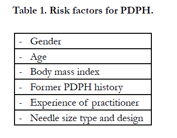
Risk factors for PDPH
Some factors were described for PDPH (Table 1). Major Risk factors for PDPH include female gender, young age, low body mass index and history of prior PDPH [10].

Female gender is blamed as an important risk factor for PDPH [8, 10, 11]. However, it was stated that this is not wholly true and it hasn’t be supported with clinical study. As the female patients is more exposed to neuroaxial block the incidence of PDPH seem higher in this population [6].
The patients between 18-30 years old are inclined to PDPH and PDPH incidence is decreased in elderly patients [6, 10, 12-15]. It was state that PDPH incidence is higher especially in young female patients due to increased fiber elasticity of dura mater [12].
Increased BMI are related to lower incidence of PDPH [10, 13, 16]. Higher intraabdominal pressure leads to increased pressure on dependent epidural vein. Thus, this event causes a protection to CSF leakage through dural puncture.
It was stated that PDPH likelihood is increased in patients experienced PDPH formerly [10, 13].
Patients with chronic headaches were reported to be more likely to PDPH.
The experience of the users is very important in regional anesthesia or dural puncture for CSF analyzing. The intervention can be done successfully with more less attempt in skilled hands. Increased attempt and ponction are related to increased PDPH likelihood. Accidental dural puncture during epidural intervention is also related to increased PDPH likelihood [9].
Needle size, type and design have become essential research on studies aimed to investigate PDPH. The radius and sharpness of the needle, the place of the hole upon the needle etc were investigated since the first neuroaxial block.
Quincke type needle has cutter surface and midline hole. It is generally known as a standard needle and has widespread use [8]. It was stated that the sharpness surface of the Quincke needle cut the dural fiber during the neuroaxial intervention and lead to more CSF leakage. However, it was also stated that blunt ended needle doesn’t cause trauma on the dural fiber and then lead to more less PDPH [17].
Pencil point needle was produced and presented for use in the later years. There two type atraumatic pencil point needle in the use nowadays: Whitacre and Sprotte. The tip of the Whitacre needle is diamond shaped. The hole of the needle is located 1mm from the tip of the needle. The tip of the Sprotte needle is conical, however, the other properties of this needle is similar with Whitacre type needle. As the hole of pencil point needle located behind the tip of the needle, it is needed to push forward for obtain CSF. It was reported that pencil point needle cause paresthesia arising from neuronal damage [18]. Therefore, atraucan needle was introduced to use. The hole of the needle is located the tip of the needle.
PDPH incidence may vary according to needle type. The widespread opinion is PDPH incidence is less the others. It was stated that as the pencil point needle has not transvers tip it isn’t cut the longitudinal dural fibers [19, 20]. Ross et al., reported that PDPH incidence with the use of Sprotte needle is less frequently seen than those with Quincke needle [21]. Similarly, Buettner et al., stated at the end of their study that PDPH incidence with the use of Whitacre needle is less frequently seen than those with Quincke needle [22]. Santanen et al., reported that PDPH incidence with the use of 27 gauge Whitacre needle is less frequently seen than those with the same radius Quincke needle [23].
Despite the presence of a number of publications reporting that pencil point needles causes less PDPH because the needles did not cut dural fibers, Reina et al., examined the fibers with microscopic dissection in cadavers and reported that the dural fibers were not place longitudinally and parallel [24]. It was demonstrated that pencil point needles performed more traumatic file than quincke needles’ in electron microscopic investigation [25]. The less incidence of PDPH with pencil point needles despite greater trauma has been explained by the fact that the pencil point needles leads to irregularly tear in the dura mater causing inflammatory reaction that prevents the CSF leakage.
One of the most important factors affecting the incidence of PDPH is the diameter of the needle used. The PDPH incidence is increase when the larger needle is used [7, 8, 26]. Kang et al., found less PDPH with smaller gauge needle in their study compared 26 and 27-gauge Quincke needles [27]. On the other hand it was stated that the success rate of neuroaxial block is decreased due to technical problems [28, 29].
The aspect of the needle may affect incidence of the PDPH. It was stated that the incidence is less encountered by paramedian approach [19, 30, 31]. Moreover, Flaatten et al., found that PDPH risk is decreased when the needle is used parallel to longitudinally axis of vertebral column [32].
Clinic Course of PDPH
PDPH is generally occurred in a week after the intervention, although it may be occurred in time letter than expected [33]. PDPH is described as an obtuse pain in localized generally frontal and occipital region of the head, less often in neck and shoulder. Nausea, vomiting, visual and aural disorders, dizziness and neck stiffness may accompany with PDPH. All of these symptoms are generally disappeared within two weeks. The International Headache Society described PDPH as a bilateral pain started within 7 days after the intervention and disappeared within 14 days [5]. The headache typically worsens within the 15 min of assuming the upright position, improves within 30 min of resuming the recumbent position [5, 33].
Treatment of PDPH
Treatment of PDPH is generally easy despite severe clinical course. It was stated that PDPH can be disappeared within six weeks without medical treatment [34, 35]. However, increased consciousness about PDPH makes possible raised patients be admitted to hospital for treatment.
Symptomatic and supportive treatment is mostly first choice for PDPH. Bed rest is described as a “sine qua non” among these supportive therapies. The aim of the bed rest is to reduce CSF leakage via dural puncture arising from gravity. However, it was asserted that bed rest isn’t serve a healing for PDPH patients [36, 37]. Moreover, it was claimed that bed rest after the surgical operation lead to worsening in special patients group [38]. Therefore, bed rest was described as supportive, not protective, therapy after occurring the PDPH [13]. Another paramedical therapy is about strict abdominal roller bandage and patient’s position.
Intra abdominal pressure is decanted to epidural space via epidural vein related abdominal vein with the use of strict abdominal roller bandage. Thus, increased epidural pressure reduces the CSF leakage leading to attenuate the PDPH [39]. Similar mechanism is deemed for prone position, however, this theory hasn’t been confirmed with clinical study [40].
The aim of medical treatment is completion of reduced volume of CSF, prevention of secondary cerebral vasodilatation arising from CSF leakage and restoration of dural puncture. Orally or intravenous fluid replacement are good for patients with PDPH. It is thought that increased intravenous volume enable more production of CSF.
The aim of this treatment is to increase intravenous volume enabling produce CSF. Isotonic saline or the other simple crystalloids are the most using fluids. Gelatin or starch based colloid are alternative option for intravenous fluid treatment of PDPH.
It was demonstrated that caffeine cause vasoconstriction of intracerebral vessels and reduce cerebral blood flow and the attenuate of PDPH [41-43]. Similarly, sumatriptan, 5-HT receptor antagonist, also provide benefit with the same mechanism for patients with PDPH [44-46]. On the other hand, it was stated that methyl ergonovine [47], cosyntropine [48] and dexamethasone [49] may be helpful for PDPH.
This technic was described a half century ago [50]. The patient’s own blood is obtained from the patient’s arm and then it is injected to epidural space on the same epidural level. Thus, clotting the blood in front of the dural puncture prevent CSF leakage. The amount of the blood should be 20-30 ml approximately [51].
Epidural fluid can be used as an alternative treatment if autologous blood patch is contraindicated [52, 53]. As the colloid fluid is more viscos, reabsorbtion of it from the epidural space is delayed. It causes a greater and longer compression, enabling closure of the dural tap [54].
Conclusion
As the PDPH is one of the most frequently complication of the neuroaxial intervention and make worse the patient’s clinical condition, it should be realized and these patients should be monitored closely.
References
- Wulf HF (1998) The centennial of spinal anesthesia. Anesthesiology. 89(2): 500-6.
- Uyar Türkyilmaz E, Camgöz Eryilmaz N, Aydin Güzey N, Moraloğlu Ö (2016) Bilateral greater occipital nerve block for treatment of post-dural puncture headache after caesarean operations. Braz J Anesthesiol. 66(5): 445-450.
- Castrillo A, Tabernero C, García-Olmos LM, Gil C, Gutiérrez R, et al., (2015) Postdural puncture headache: impact of needle type, a randomized trial. Spine J. 15(7): 1571-1576.
- Lee JA (1979) Arthur Edward James Barker 1850-1916. British pioneer of regional analgesia. Anaesthesia. 34(9): 885-891.
- Evans RW, Armon C, Frohman EM, Goodin DS (2000) Assessment: prevention of post-lumbar puncture headaches: report of the therapeutics and technology assessment subcommittee of the american academy of neurology. Neurology. 55(7): 909-914.
- Gaiser R (2006) Postdural puncture headache. Curr Opin Anaesthesiol. 19(3): 249-253.
- Choi PT, Galinski SE, Takeuchi L, Lucas S, Tamayo C, et al., (2003) PDPH is a common complication of neuraxial blockade in parturients: a meta-analysis of obstetrical studies. Can J Anaesth. 50(5): 460-469.
- Turnbull DK, Shepherd DB (2003) Post-dural puncture headache: pathogenesis, prevention and treatment. Br J Anaesth. 91(5): 718-29.
- Reynolds F (1993) Dural puncture and headache. BMJ. 306: 874-876.
- Bezov D, Lipton RB, Ashina S (2010) Post-dural puncture headache: part I diagnosis, epidemiology, etiology, and pathophysiology. Headache. 50(7):1144-52.
- Wu CL, Rowlingson AJ, Cohen SR, Michaels RK, Courpas GE, et al., (2006) Gender and post-dural puncture headache. Anesthesiology. 105(3): 613-618.
- Evans RW (1998) Complications of lumbar puncture. Neurol Clin. 16(1): 83-105.
- Ghaleb A, Khorasani A, Mangar D (2012) Post-dural puncture headache. Int J Gen Med. 5: 45-51.
- Flaatten H, Rodt S, Rosland J, Vamnes J (1987) Postoperative headache in young patients after spinal anaesthesia. Anaesthesia. 42(2): 202-205.
- Rasmussen BS, Blom L, Hansen P, Mikkelsen SS (1989) Postspinal headache in young and elderly patients. Two randomised, double-blind studies that compare 20- and 25-gauge needles. Anaesthesia. 44(7): 571-573.
- Liu S, Carpenter RL, Neal JM (1995) Epidural anesthesia and analgesia. Their role in postoperative outcome. Anesthesiology. 82(6): 1474-1506.
- Grene HM (1926) Lumbar puncture and the prevention of post puncture headache. JAMA. 86(6): 391-392.
- Sharma SK, Gambling DR, Joshi GP, Sidawi JE, Herrera ER (1995) Comparison of 26-gauge Atraucan and 25-gauge Whitacre needles: insertion characteristics and complications. Can J Anaesth. 42(8): 706-710.
- Norris MC, Leighton BL, DeSimone CA (1989) Needle bevel direction and headache after inadvertent dural puncture. Anesthesiology. 70(5): 729-731.
- Vallejo MC, Mandell GL, Sabo DP, Ramanathan S (2000) Postdural puncture headache: a randomized comparison of five spinal needles in obstetric patients. Anesth Analg. 91(4): 916-920.
- Ross BK, Chadwick HS, Mancuso JJ, Benedetti C (1992) Sprotte needle for obstetric anesthesia: decreased incidence of post dural puncture headache. Reg Anesth. 17(1): 29-33.
- Buettner J, Wresch KP, Klose R (1993) Postdural puncture headache: comparison of 25-gauge Whitacre and Quincke needles. Reg Anesth. 18(3): 166- 169.
- Santanen U, Rautoma P, Luurila H, Erkola O, Pere P (2004) Comparison of 27-gauge (0.41-mm) Whitacre and Quincke spinal needles with respect to post-dural puncture headache and non-dural puncture headache. ActaAnaesthesiol Scand. 48(4): 474-479.
- Reina MA, Dittmann M, López Garcia A, van Zundert A (1997) New perspectives in the microscopic structure of human dura mater in the dorsolumbar region. Reg Anesth. 22(2): 161-166.
- Reina MA, de Leon-Casasola OA, Lopez A, De Andres J, Martin S, et al., (2000) An in vitro study of dural lesions produced by 25-gauge Quincke and Whitacre needles evaluated by scanning electron microscopy. Reg Anesthzain Med. 25(4): 393-402.
- Lambert DH, Hurley RJ, Hertwig L, Datta S (1997) Role of needle gauge and tip configuration in the production of lumbar puncture headache. Reg Anesth. 22(1): 66-72.
- Kang SB, Goodnough DE, Lee YK, Olson RA, Borshoff JA, et al., (1992) Comparison of 26- and 27-G needles for spinal anesthesia for ambulatory surgery patients. Anesthesiology. 76(5): 734-738.
- Flaatten H, Rodt SA, Vamnes J, Rosland J, Wisborg T, et al., (1989) Postdural puncture headache. A comparison between 26- and 29-gauge needles in young patients. Anaesthesia. 44(2): 147-149.
- Hoskin MF (1998) Spinal anaesthesia--the current trend towards narrow gauge atraumatic (pencil point) needles. Case reports and review. Anaesth Intensive Care. 26(1): 96-106.
- Lybecker H, Møller JT, May O, Nielsen HK (1990) Incidence and prediction of postdural puncture headache. A prospective study of 1021 spinal anesthesias. Anesth Analg. 70(4): 389-394.
- Ready LB, Cuplin S, Haschke RH, Nessly M (1989) Spinal needle determinants of rate of transdural fluid leak. Anesth Analg. 69(4): 457-460.
- Flaatten H, Thorsen T, Askeland B, Finne M, Rosland J, et al., (1998) Puncture technique and postural postdural puncture headache. A randomised, double-blind study comparing transverse and parallel puncture. Acta Anaesthesiol Scand. 42(10): 1209-1214.
- Porhomayon J, Zadeii G, Yarahamadi A, Nader ND (2013) A case of prolonged delayed postdural puncture headache in a patient with multiple sclerosis exacerbated by air travel. Case Rep Anesthesiol. 2013: 253218.
- Vandam LD, Dripps RD (1956) Long-term follow-up of patients who received 10,098 spinal anesthetics; syndrome of decreased intracranial pressure (headache and ocular and auditory difficulties). J Am Med Assoc. 161(7):586-591.
- Costigan SN, Sprigge JS (1996) Dural puncture: the patients' perspective. A patient survey of cases at a DGH maternity unit 1983-1993. Acta Anaesthesiol Scand. 40(6): 710-714.
- Thoennissen J, Herkner H, Lang W, Domanovits H, Laggner AN, et al., (2001) Does bed rest after cervical or lumbar puncture prevent headache? A systematic review and meta-analysis. CMAJ. 165(10): 1311-1316.
- Spriggs DA, Burn DJ, French J, Cartlidge NE, Bates D (1992) Is bed rest useful after diagnostic lumbar puncture? Postgrad Med J. 68(801): 581-583.
- Fassoulaki A, Sarantopoulos C, Andreopoulou K (1991) Is early mobilization associated with lower incidence of postspinal headache? A controlled trial in 69 urologic patients. Anaesthesiol Reanim. 16(6): 375-378.
- Mosavy SH, Shafei M (1975) Prevention of headache consequent upon dural puncture in obstetric patient. Anaesthesia. 30(6): 807-809.
- Handler CE, Smith FR, Perkin GD, Rose FC (1982) Posture and lumbar puncture headache: a controlled trial in 50 patients. J R Soc Med. 75(6): 404-407.
- Camann WR, Murray RS, Mushlin PS, Lambert DH (1990) Effects of oral caffeine on postdural puncture headache. A double-blind, placebo-controlled trial. Anesth Analg. 70(2): 181-184.
- Yücel A, Ozyalçin S, Talu GK, Yücel EC, Erdine S (1999) Intravenous administration of caffeine sodium benzoate for postdural puncture headache. Reg Anesth Pain Med. 24(1): 51-54.
- Shenkin HA, Novack P (1954) Clinical implications of recent studies on cerebral circulation of man. AMA Arch Neurol Psychiatry. 71(2): 148-159.
- Hodgson C, Roitberg-Henry A (1997) The use of sumatriptan in the treatment of postdural puncture headache. Anaesthesia. 52(8): 808.
- Connelly NR, Parker RK, Rahimi A, Gibson CS (2000) Sumatriptan in patients with postdural puncture headache. Headache. 40(4): 316-319.
- Carp H, Singh PJ, Vadhera R, Jayaram A (1994) Effects of the serotoninreceptor agonist sumatriptan on postdural puncture headache: report of six cases. Anesth Analg. 79(1): 180-182.
- Hakim S, Khan RM, Maroof M, Usmani H, Huda W, et al., (2005) Methylergonovine maleate (methergine) relieves postdural puncture headache in obstetric patients. Acta Obstet Gynecol Scand. 84(1): 100.
- Carter BL, Pasupuleti R (2000) Use of intravenous cosyntropin in the treatment of postdural puncture headache. Anesthesiology. 92(1): 272-274.
- Hamzei A, Basiri-Moghadam M, Pasban-Noghabi S (2012) Effect of dexamethasone on incidence of headache after spinal anesthesia in cesarean section. A single blind randomized controlled trial. Saudi Med J. 33(9): 948-953.
- Gormley JB (1960) Treatment of post-spinal headache. Anesthesiology. 21: 565-556.
- Crawford JS (1980) Experiences with epidural blood patch. Anaesthesia. 35(5): 513-515.
- Souron V, Hamza J (1999) Treatment of postdural puncture headaches with colloid solutions: an alternative to epidural blood patch. Anesth Analg. 89(5): 1333-1334.
- Che X, Zhang W, Xu M (2016) Continuous epidural pumping of saline contributes to prevent and treat postdural puncture headache. J Clin Anesth. 34: 154-158.
- Salvador L, Carrero E, Castillo J, Villalonga A, Nalda MA (1992) Prevention of post dural puncture headache with epidural-administered dextran 40. Reg Anesth. 17(6): 357–358.


