Immature Coconut Water: A Renal Protective Agent in Wistar Rats
Okafor CW1, Ekezie J1*, Ndubuka GI2, Ezejiofor, T.I.N3
1 Department of Biomedical Technology, School of Health Technology, Federal University of Technology, Owerri, Nigeria.
2 Department of Anatomy, School of Health Technology,Federal University of Technology, Owerri, Nigeria.
3 Department of Biotechnology, School of Biological Sciences, Federal University of Technology, Owerri, Nigeria.
*Corresponding Author
Ekezie Jervas,
Department of Anatomy, Federal University of Technology,
P.M.B 1526 Owerri Nigeria.
Tel: +234 806 5430037
E-mail ekeziejervas@gmail.com
Received: February 29, 2016; Accepted: March 16, 2016; Published: March 30, 2016
Citation: Okafor CW, Ekezie J, Ndubuka GI, Ndubuisi T (2016) Immature Coconut Water: A Renal Protective Agent in Wistar Rats. Int J Anat Appl Physiol. 2(2), 20-25.doi: dx.doi.org/10.19070/2572-7451-160004
Copyright: Ekezie J© 2016. This is an open-access article distributed under the terms of the Creative Commons Attribution License, which permits unrestricted use, distribution and reproduction in any medium, provided the original author and source are credited.
Abstract
The biochemical characteristics of immature coconut water (ICW) have presented it as a therapeutic agent that can be used for preventive, management and curative purposes. Development of a more efficient and cost effective means of prevention and management of kidney disorders through antioxidant activities were the objectives for embarking on the current experiment. This experiment is aimed at investigating the protective effect of ICW against carbon tetrachloride (CCl4) induced renal toxicity. Twenty(20) rats were fed on standard diet and divided into four groups. Rats in group 1 and group 2 were injected intraperitoneally (i.p) with olive oil. Group 1 received tap water only, while group 2 received ICW (100ml/kg body weight/day) only. Rats in Group 3 and Group 4 were injected i.p with CCl4 (5ml/kg body weight). Group 3 received tap water, while group 4 received ICW (100ml/kg body weight/day) only. At the end of the experiment (1 week), blood was collected for biochemical analysis. The present findings revealed that, CCl4 decreased theserum level of kidney markers and increased the serum levels of some electrolytes; but these effects were prevented by the prior administration ICW on rats. The present study concluded that ICW administration played a protective role against CCl4-induced kidney damages in Wister rats. These protective effects were in the form of improving of kidney markers and electrolyte activities, in CCl4- intoxicated rats. In the future, a dose dependent protective effect, and in vitro and in vivo regenerative effect of ICW on the kidney could be investigated.
2.Introduction
3.Materials and Methods
3.1.Plant Materials
3.2.Animal
3.3.Chemical
3.4.Induction of renal totoxicity by CCl4
3.5.Determination of Dosage for the Administration of Inmature Coconut Water (ICW) in Experimental Animals.
3.6.Experimental Group and Protocol
3.7.Collection of Blood Samples
3.8.Biochemical Techniques
3.8.Statistical Data Analysis
4.Results and Discussion
5.Conclusion & Recommendation
6.References
KeyWords
Protection; Coconut water; Serum; Kidney; Biochemistry.
Introduction
The abundance of polyunsaturated fatty acids and the presence of cytochrome P450 makes the kidney an organ particularly vulnerable to reactive oxygen species (ROS) attack [1-3]. The concentration of ROS reaches its peak <1-6 hours, depending on exposure and concentration or dose [4-8]. Carbon tetrachloride toxicity is dependent on the excessive production of the trichloromethyl radical (CCl3) [9]. Early studies showed that free radicals, such as trichloromethyl (CCl3) and oxygen-centered lipid radicals (LO or LOO, or both), are generated during CCl4 metabolism by hepatic cellular cytochrome P450 [10].
A common manifestation and a mediator of cardiovascular, neurological, and numerous other complications of renal failure is oxidative stress [3]. It constitutes the mechanisms of production and progression of numerous renal diseases [11], as it mediates a wide range of renal impairments, from acute renal failure [12-16],rhabdomyolysis [12, 17] obstructive nephropathy [18, 19], hyperlipidemia [18, 20-22] and glomerular damage [23- 25] to chronic renal failure and hemodialysis [26-29].
Despite the well-developed antioxidant systems to neutralize most detrimental effects of these oxidizing species in living organisms, a continuous production of oxidizing agent would eventually overwhelm it [30]. Therefore an antioxidizing action induced by antioxidizingagent would be vital in protecting against CCl4- induced damage in the kidney [31].
The antioxiding action of Immature coconut water (ICW) has been reported [30-34], and it was observed to be more potent in fresh coconut water samples than in heated acid or alkali treated samples [35]. The protective effect of ICW against toxins has been studied, and its protective effect is evident from the histopathological studies of the liver and kidney in ICW treated Wister rats, which did not show any distorted sinusoid and fatty infiltrations or necrosis, as observed in CCl4-intoxicated rats [36].
In this present study, we investigated the protective effects of ICW against CCl4-induced kidney toxicity in rats, by examining serum levels of kidney markers and some electrolytes.
Materials and Methods
Young coconuts were (Cocosnucifera L.) collected from Eziobodo Community, in Owerri West, Imo State of Nigeria. It was authenticated and identified the department of Forestry & Wildlife, School of Agricultural Technology, Federal University of Technology, Owerri; as a dwarf (autogamous) Coconut (Cocosnucifera L. Arecaceae). The fresh immature coconut water (ICW) was obtained from the coconuts each time it is required for administration on the Wister rats.
A total of 20 adult male Wistar rats with body weights ranging from 175g to 200g obtained from Animal house of the Department of Forestry & Wildlife, School of Agricultural Technology, Federal University of Technology, Owerri, Nigeria were used in the study. The animals were allowed acclimatization in the laboratory conditions for two weeks before the commencement of the study. During which, the experimental animals were housed in cages, kept on a 12 h/12 h light/dark cycle and had free access to standard rodent pellet diet and water ad libitum. The experimental procedures adopted in this study were in strict compliance with the United States National Institutes of Health Guidelines for Care and Use of Laboratory Animals in Biomedical Research [37].
Carbon tetrachloride (Riedel-de Haen AG Seelze-Hannover), Olive oil and other chemicals and solvents were of highest grade commercially available.
Renal toxicity was induced by intraperitoneal injection of Carbon tetrachloride CCl4, diluted with distilled water and vector (Olive oil) in the ratio of 1:2:0.5 respectfully. Dosage was determined using 5ml/kg body weight, as a standard. Therefore, the specific dosage for each Wister rat was calculated thus:
Milligram Equivalent for renal toxicity induction = (5ml×weight of rats (g))/ 1000g
Immature coconut water (ICW) was administered through intragastric injection. The dosage was determined using 100ml/kg body weight, as a standard. Therefore the specific dosage for each Wister rat was calculated thus:
Milligram Equivalent for ICW Administration = (100ml×weight of rat (g))/ 1000g
The rats were divided randomly into 4 groups comprising of 5 rats in each group. They were all fed with the same diet throughout the experimental period. The experimental design is described as follows:
This group is made up of 5 male rats with weights ranging from 175g-200g. Rats were fed with rodent pellets and tap water (basal diet), and were injected intraperitoneally on the 7th day of the experiment with olive oil (0.5ml/kg body weight) only.
This group is made up of 5 male rats with weights ranging from 175g-200g. Rats were fed with rodent pellet, received ICW (100 ml/kg body weight/day) as their sole source of drinking water, and were injected intraperitoneally on the 7th day of the experiment with olive oil (0.5ml/kg body weight). This group served as positive control. The calculated dosage ICW was given in fragments of 3 times (i.e. 8am, 1pm, and 5pm) daily; via intragastric injection.
This group is made up of 5 male rats with weights ranging from 175g-200g. Rats were fed basal diet and tap water, and then they were intoxicated via intraperitoneal injection on the 7th day of the experiment with CCl4 diluted with distilled water and Olive oil, at a ratio of 1:2:0.5 respectively. The dosage given was 5ml/kg body weight.
This group is made up of 5 male rats with weights ranging from 175g-200g. Rats were fed basal diet and ICW (100 ml/kg body weight/day) as their sole source of drinking water [the calculated dosage of ICW was given in fragments of 3 times (i.e 8am, 1pm, and 5pm) daily; via intragastric injection], and then they were intoxicated via intraperitoneal injection on the 7th day of the experiment with CCl4 diluted with distilled water and Olive oil, at a ratio of 1:2:0.5 respectively (the calculated dosage given was 5ml/kg body weight).
At the end of the experiment, the animals were fasted overnight prior to the collection of samples. Blood was obtained from the control and experimental animals through Ocular puncture, using capillary tubes. 5 ml of blood samples were collected. The blood obtained was put into EDTA containers and plain blood sample containers. Serum was obtained from the blood and used for analysis.
Commercially obtained diagnostic kits were used to determine the serum levels of biochemical parameters, as follows: Randox Laboratories Limited were used for determination of serum urea and creatinine. Teco Diagnostics were used for determination of serum bicarbonate, chloride, potassium and Sodium. Concentration of the biochemical constituents were calculated according to the manufacturers’ instructions, a standard protocol. The values obtained were used to analyze the biochemical condition of the experimental animals.
The Biochemical Parameters were analyzed using SPSS. Analysis of variance (ANOVA) was first performed. Then a post-hoc test via Bonferronifor multiple comparisons was carried out. The Biochemical Parameters wereconsidered statistically significant at p<0.05 and Confidence interval (CI) void of zero value.
Results and Discussion
The result of the biochemical activities of the wistar rat are shown in the tables that follow.
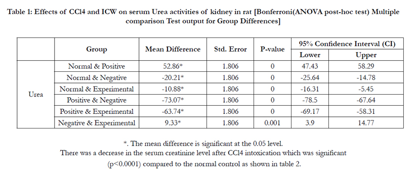
The decrease in the serum urea level was found to be significant (p<0.0001) compared to the normal control as shown in table 1.

There was a decrease in the serum creatinine level after CCl4 intoxication which was significant (p<0.0001) compared to the normal control as shown in Table 2.
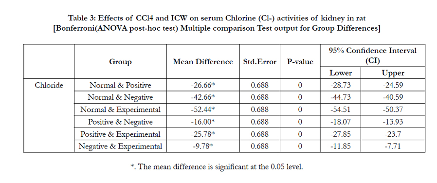
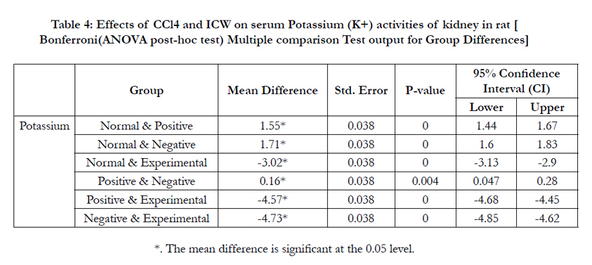
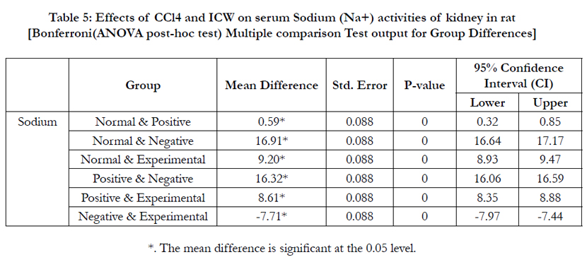
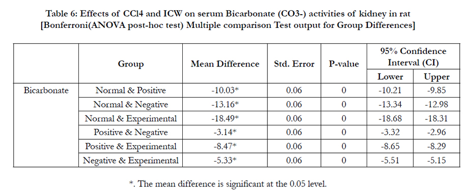
In this study also, bicarbonate (HCO3-), chloride (Cl-), potassium (K+) and Sodium (Na2+) levels in the negative control, were significantly elevated (p<0.0001) respectively, compared to the normal control group (Table 3-6).
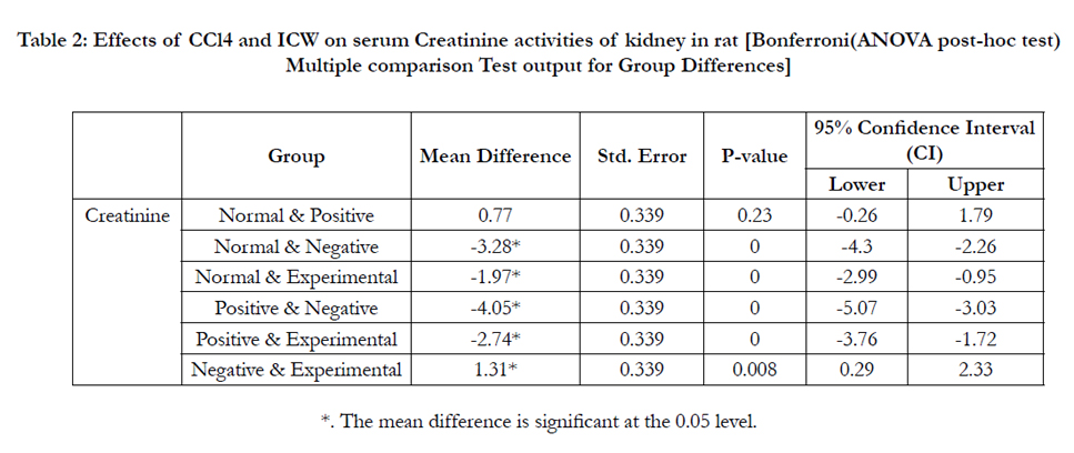
In this study, carbon tetrachloride (CCl4) treatment of Wister rats in the negative control resulted in decreased serum level of urea. The decrease in serum urea level was found to be significant (p<0.05) compared to the normal control as shown in table 1. This finding tends to support the reports obtained by previous experimental studies [38, 39]. Also, the reports on the effect of CCl4 on serum creatinine level tends be contradicting among some investigators. While some investigators [39-42] found no significant effect of CCl4 on serum creatinine level, our present study follows the report of other investigators [43] which observed a decrease in the serum creatinine level after CCl4 intoxication which was significant (p<0.05) compared to the normal control as shown in table 2.
These alterations can be attributed to the disruption of the permeability of plasma, lysosomal, and mitochondrial membranes, as a result of the release of highly reactive oxides formed by the metabolism of CCl4 [44, 45]. However, these trends were reverted in the experimental group as the serum urea and creatinine levels increase significantly. Elevation of these waste products in the kidney as depicted in table 1 & 2 are an indication of maintenance of renal function [46].
Maintenance of osmotic balance of the blood is done by reabsorption of ions, and it is one of the principal functions of the kidney [47]. In this study, bicarbonate (HCO3-), chloride (Cl-), potassium (K+) and Sodium (Na2+) levels in the negative control, were elevated significantly (p<0.05) compared to the normal control group (table 3-6). The significant increase in Na2+ concentration may indicate excess destruction of cells [47]. The serum increase of electrolytes is further evidence of renal impairment [48]. Although, a keen observation of the different levels of elevations show that they tend to vary among the various groups.
These renal protective effects were manifestations of antioxidant properties of the micronutrients contained in ICW. This is consistent with the report that micronutrients act directly to quench free radicals by donating electrons, or indirectly as a part of metallo enzymes [30].
Conclusion & Recommendation
It is considerable enough from the results of this study that ICW was able to suppress effectively, the effects of the radical oxidizing species produced from CCl4 metabolism. This it achieved by exhibiting antioxidant activities.
There is need for an in vitro and an in vivo investigation into the curative and regenerative effect of young coconut water on renal cells.
References
- Tomasi A, Albano E, Banni S, Botti B, Corongiu F, et al. (1987) Free-radical metabolism of carbon tetrachloride in rat liver mitochondria. A study of the mechanism of activation. Biochem J 246(2): 313-317.
- Reinke LA, Janzen EG (1991) Detection of spin adducts in blood after administration of carbon tetrachloride to rats. Chem Biol Interact 78(2): 155-165.
- Ichikawa I, Kiyama S, Yoshioka T (1994) Renal antioxidant enzymes: their regulation and function. Kidney Int 45(1): 1-9.
- McCay PB, Lai EK, Poyer JL, DuBose CM, Janzen EG (1984) Oxygen- and carbon-centered free radical formation during carbon tetrachloride metabolism. Observations of lipid radicals in vivo and in vitro. J Biol Chem 259(4): 2135-2143.
- Mico BA, Pohl LR (1983) Reductive oxygenation of carbon tetrachloride. Trichloromethylperoxyl radical as a possible intermediate in the conversion of carbon tetrachloride to electrophilic chlorine. Arch Biochem Biophys 225(2): 596-609.
- Slater TF (1982) Activation of carbon tetrachloride: chemical principles and biological significance. In Free radicals, lipid peroxidation and cancer. Academic Press, New York. 243-274.
- Poyer JL, McCay PB, Lai EK, Janzen EG, Davis ER (1980) Confirmation of assignment of the trichloromethyl radical spin adduct detected by spin trapping during 13C-carbon tetrachloride metabolism in vitro and in vivo. Biochem Biophys Res Commun 94(4): 1154-1160.
- Lai EK, McCay PB, Noguchi T, Fong KL (1979) In vivo spin-trapping of trichloromethyl radicals formed from carbon tetrachloride. Biochem Pharmacol 28(14): 2231-2235.
- Behar-Cohen FF, Heydolph S, Faure V, Droy-Lefaix MT, Courtois Y, et al. (1996) Peroxynitrite cytotoxicity on bovine retinal pigmented epithelial cells in culture. Biochem Biophys Res Commun 226(3): 842-849.
- Smuckler EA, Iseri OA, Benditt EP (1962) An intracellular defect in protein synthesis induced by carbon tetrachloride. J Exp Med 116: 55-72.
- Martin-Mateo MC, Sanchez-Portugal M, Iglesias S, de Paula A, Bustamante J (1999) Oxidative stress in chronic renal failure. Ren Fail 21(2): 155-167.
- Zager RA, Burkhart K (1997) Myoglobin toxicity in proximal human kidney cells: roles of Fe, Ca2+, H2O2, and terminal mitochondrial electron transport. Kidney Int 51(3): 728-738.
- Paller MS, Weber K, Patten M (1998) Nitric oxide-mediated renal epithelial cell injury during hypoxia and reoxygenation. Ren Fail 20(3): 459-469.
- Baliga R, Ueda N, Walker PD, Shah SV (1999) Oxidant mechanism in toxic acute renal failure. Drug Metab Rev 31(4): 971-997.
- Barrouillet MP, Moiret A, Cambar J (1999) Protective effects of polyphenols against cadmium-induced glomerular mesangial cell myocontracture. Arch Toxicol 73(8-9): 485-488.
- Shah S (2001) Role of iron in progressive renal disease. Am J Kidney Dis 37(1 Suppl 2): S30-S33.
- Vanholder R, Sever MS, Erek E, Lameire N (2000) Rhabdomyolysis. J Am Soc Nephrol 11(8): 1553-1561.
- Greiber S, Kramer-Guth A, Pavenstadt H, Gutenkunst M, Schollmeyer P, et al. (1996) Effects of lipoprotein(a) on mesangial cell proliferation and viability. Nephrol Dial Transplant 11(5): 778-785.
- Klahr S (1998) Nephrology forum: obstructive nephropathy. Kidney Int 54: 286-300.
- Sakatsume M, Kadomura M, Sakata I, Imai N, Kondo D, et al. (2001) Novel glomerular lipoprotein deposits associated with apolipoprotein E2 homozygosity. Kidney Int 59(5): 1911-1918.
- Wanner C, Greiber S, Kramer-Guth A, Heinloth A, Galle J (1997) Lipids and progression of renal disease: role of modified low density lipoprotein and lipoprotein(a). Kidney Int 52(Suppl 63): S102-S106.
- Scheuer H, Gwinner W, Hohbach J, Grone EF, Brandes RP, et al. (2000) Oxidant stress in hyperlipidemia-induced renal damage. Am J Physiol 278(1): F63-F74.
- Hahn S, Krieg RJ Jr, Hisano S, Chan W, Kuemmerle NB, et al. (1999) Vitamin E suppresses oxidative stress and glomerulosclerosis in rat remnant kidney. Pediatr Nephrol 13(3): 195-198.
- Kitamura M, Ishikawa Y (1999) Oxidant-induced apoptosis of glomerular cells: intracellular signalling and its intervention by bioflavonoid. Kidney Int 56(4): 1223-1229.
- Moreno-Manzano V, Ishikawa Y, Lucio-Czana J, Kitamura M (2000) Selective involvement of superoxide anion, but not down-stream compounds hydrogen peroxide and peroxynitrite, in tumor necrosis factor-induced apoptosis of rat mesangial cells. J Biol Chem 275: 12684-12691.
- Fiorillo C, Oliviero C, Rizzuti G, Nediani C, Pacini A, et al. (1998) Oxidative stress and antioxidant defenses in renal patients receiving regular haemodialysis. Clin Chem Lab Med 36(3): 149-153.
- Fryer MJ (1997) Vitamin E may slow kidney failure owing to oxidative stress. Redox Rep 3(5-6): 259-261.
- Handelman GJ, Walter MF, Adhikarla R, Gross J, Dallal GE, et al. (2001) Elevated plasma F2-isoprostanes in patients on long-term hemodialysis. Kidney Int 59(5): 1960-1966.
- Ozaki M, Yamada Y, Matoba K, Otani H, Mune M, et al. (1999) Phospholipase A2 activity in ox-LDL-stimulated mesangial cells and modulation by alpha-tocopherol. Kidney Int Suppl 71: S171–S173.
- Shenkin A (2006) The key role of micronutrients. Clin Nutr 25(1): 1-13.
- Gideon IN, Wilson CO, Ekezie J, Iwuji SC, Okeke CU, et al. (2015) Protective Effect of Immature Coconut Water on Hepatocytes against Carbon tetrachloride-induced Liver Damage in Wister Rats. International Journal of Science and Research (IJSR) Vol. 4 Issue 12. ISSN (Online): 2319-7064,
- Mantena SK, Jagadish, Badduri SR, Siripurapu KB, Unnikrishnan MK (2003) In vitro evaluation of antioxidation properties of Cocos nucifera Linn. Water. Nahrung 47(2): 126-131.
- Evans P, Halliwell B (2001) Micronutrients: Oxidant/antioxidant Status. Br J Nutr 85(Suppl 2): S67-S74.
- Nevin KG, Rajamohan T (2006) Virgin coconut oil supplemented diet increases the antioxidant status in rats. Food Chem 99(2): 260-266.
- Prades A, Dornier M, Diop N, Jean-Pierre P (2012) Coconut water uses, composition and properties: a review. Fruits 67(2): 87-107.
- Loki AL, Rajamohan T (2003) Hepatoprotective and antioxidant effect of tender coconut water on CCl4 induced liver injury in rats. Indian J Biochem Biophy 40(5): 354-357.
- United States Department of Agriculture (USDA) (2008) Nuts, coconut water. National nutrient database for standard reference.
- Sreepriya M, Devaki T, Nayeem M (2001) Protective effects of Indigofera tinctoria L. against D-Galactosamine and carbon tetrachloride challenge on ‘in situ’ perfused rat liver. Indian J Physiol Pharmacol 45(4): 428-434.
- Khan AA, Alzohairy M (2011) Hepatoprotective effects of camel milk against CCl4-induced hepatotoxicity in Rats. Asian J Biochem 6(2): 171-180.
- Palaparthy R, Kastrissios H, Gulati A (2001) Pharmacokinetics of diaspirin cross-linked haemoglobin in a rat model of hepatic cirrhosis. J Pharm Pharmacol 53(2): 179-185.
- Wirth KJ, Bickel M, Hropot M, Günzler V, Heitsch H, et al. (1997) The bradykinin B2 receptor antagonist icatibant (HOE 140) corrects avid Na+ retention in rats with CCl4-induced liver cirrhosis: possible role of enhanced microvascular leakage. Eur J Pharmacol 337(1): 45-53.
- Ozdogan O, Goren MZ, Ratip S, Giral A, Moini H, et al. (2002) Role of endothelin-1 in a cirrhotic rat model with endotoxin induced acute renal failure. Hepatol Res 24(2): 114-124.
- Cruz C, Ibarra-Rubio ME, Pedraza-Chaverri J (1993) Circulating levels of active, total and inactive renin (prorenin), angiotensin I and angiotensinogen in carbon tetrachloride-treated rats. Clin Exp Pharmacol Physiol 20(2): 83-88.
- Shi J, Aisaki K, Ikawa Y, Wake K (1998) Evidence of hepatocyte apoptosis in rat liver after the administration of carbon tetrachloride. Am J Pathol 153(2): 515-525.
- Manibusan MK, Odin M, Eastmond DA (2007) Postulated carbon tetrachloride mode of action: a review. J Environ Sci Health C Environ Carcinog Ecotoxicol Rev 25(3): 185-209.
- Orth SR, Ritz E (1998) The nephrotic syndrome. N Engl J Med 338(17): 1202-1211.
- Rose BD, Post TW (2001) Regulation of the effective circulating volume. In Clinical physiology of acid-base and electrolyte disorders. (5th edtn), McGraw Hill, New York.
- Cameron JS, Greger R (1998) Renal function and testing of function. In Oxford Textbook of Clinical Nephrology. Oxford University Press, UK. 36- 39.


