Y-STR Profiling of Semen Stain Evidences of Azoospermic Individuals
Ali M. Al-Dosary1, Ahmed M. Refaat1*, Saranya R. Babu1, Abdul Rauf Choudhry1
Naif Arab University for Security Sciences (NAUSS), Saudi Arabia.
*Corresponding Author
Ahmed M. Refaat Ph.D,
Naif Arab University for Security Sciences (NAUSS),
Saudi Arabia.
Tel: 966595229237
E-mail: dr.ahmed_mr@yahoo.com
Article Type : Research Article
Received: August 27, 2015; Accepted: November 19, 2015; Published: November 20, 2015
Citation: Ali M. Al-Dosary, Ahmed M. Refaat, Saranya R. Babu, Abdul Rauf Choudhry (2015) Y-STR Profiling of Semen Stain Evidences of Azoospermic Individuals. Int J Forensic Sci Pathol. 3(11), 210-214. doi: dx.doi.org/10.19070/2332-287X-1500050
Copyright: Ahmed M. Refaat© 2015. This is an open-access article distributed under the terms of the Creative Commons Attribution License, which permits unrestricted use, distribution and reproduction in any medium, provided the original author and source are credited.
Abstract
Background: Nowadays, STR polymorphism is the method of choice in all governmental and private laboratories working in DNA profiling, since it allows DNA investigators to analyze all types of biological evidences, besides obtaining high discrimination power for a large number of cases including both criminal identification and paternity tests as well. Azoospermia is a condition that when occurs in certain individual it causes a complete absence of the major DNA source in semen which is spermatozoa leading to infertility. The aim of this study is to obtain STR profiles from those minute DNA quantities that might be present in the semen stain evidences of Azoospermic individuals, to generate confidential results presented to the court after the analysis of these special type of biological evidences.
Methods: 100 semen stain trace samples divided into two groups (test & control samples) were recovered by a simulating manner to that which is done in the crime scene investigation, then DNA was extracted using QIAamp DNA Micro Kit, with special modifications in the extracting procedure applied for the Azoospermic test samples to concentrate and obtain minute quantities of DNA. DNA quantification was done using a Real-time PCR utilizing Quantifiler Duo Kit. PCR was done using an AmpFlSTR Y-Filer Kit, then amplified products for all test and control samples were typed using a 3130 Genetic Analyzer.
Results: Full Y-STR profiles were obtained from semen stain evidences attributed to Azoospermic individuals, with a percentage reaches 100%.
Conclusion: The methodology followed in this research should be applied in case of performing DNA typing for semen stain evidences recovered from crime scenes, to guarantee obtaining full DNA profiles even with Azoospermic samples.
2.Introduction
3.Materials and Methods
4.Results
5.Discussion
6.Conclusion
7.Acknowledgement
8.References
Keywords
DNA Profiling; Y-STRs; Semen Stain Evidences; Azoospermia.
Introduction
Genetic fingerprinting utilizing STR polymorphism is now the method of choice in all laboratories investigating biological evidences recovered from the crime scene. Y-STR profiling is an important branch of genetic Fingerprinting that links male individuals from the same paternal lineage. When this technology arose, it increases the types of forensic applications which can be solved via DNA Profiling [11]. In crime scene investigation, one of the most important biological evidences is the semen. It is found in various types of crimes as rape, pederasty, and sex-related homicides. Certain precautions must be taken by the CSI examiners in order to detect, recover, and preserve semen stain evidences until processed by the forensic laboratory experts. Semen stain evidences can be beneficial in identifying the perpetrator and/or the victim in many cases, this can be achieved through analyzing the genomic DNA contained in the semen.
Semen is an organic fluid that may contain spermatozoa. It is secreted by the gonads (sexual glands) and other sexual organs of male, and can fertilize female ova. In humans, semen contains several components besides spermatozoa: proteolytic and other enzymes as well as fructose are elements of seminal fluid which promote the survival of spermatozoa, and provide a medium through which they can move or "swim". Semen is produced and originates from the seminal vesicle, which is located in the pelvis. The process that results in the discharge of semen is called "ejaculation" [8]. During the process of ejaculation, sperm passes through the ejaculatory ducts and mixes with fluids from the seminal vesicles, the prostate, and the bulbourethral glands to form the semen. The seminal vesicles produce a yellowish viscous fluid rich in fructose and other substances that makes up about 70% of human semen. The prostatic secretion, influenced by dihydrotestosterone, is a whitish (sometimes clear), thin fluid containing proteolytic enzymes, citric acid, acid phosphatase and lipids. The bulbourethral glands secrete a clear secretion into the lumen of the urethra to lubricate it [9].
Azoospermia is the most severe form of male infertility, it is the medical condition of a man not having any measurable level of sperm in his semen. In humans, Azoospermia affects about 1% of the male population and may be seen in up to 20% of male infertility situations [10].
Our study investigates semen stain evidences similar to those found, recovered, and processed by criminal lab experts, these evidences are attributed to Azoospermic individuals - aiming to extract, concentrate, amplify, and obtain full Y-STR profiles from those minute traces of DNA present in such type of biological evidences, to help improving identification purposes regarding Azoospermic individuals.
Materials and Methods
One hundred semen stain samples from one hundred individuals previously subjected to semen analysis, were included in this study and are categorized into two groups: Group I (control group): consists of 50 semen stain samples taken from men aged from 25 to 35 years from the Saudi population after their informed consents, and it was proved from the results indicated in the report of their semen analysis that they are all having normal sperm count and charecteristics and they are all fertile persons. Group II (patients group): consists of 50 semen stain samples taken from men aged from 25 to 35 years from the Saudi population after their informed consents, and it was proved from the results indicated in the report of their semen analysis that they all have Azoospermia (sperm count = 0). A photocopy for the clinical report of semen analysis done for each individual (from both groups) was obtained from a private clinical lab in Riyadh prior to taking semen stain samples in the duration from Septemberer to March 2015. All semen samples were collected and stored at -20°C.
Samples were prepared prior to performing presumptive tests through liquifying at room temperature then votexing for 5-10 sec, then withdrawing 300 microlitres from each sample and deposit on a clean 5cm2 piece of white cloth 100% cotton. After semen deposition, semen stains were allowed to dry in room temperature for 45 minutes in a simulating manner by which semen stain evidences are found and recovered from the crime scene.
Acid phosphatase test was the first chemical test done on all semen stain samples (group I and group II) for detecting semen by the same way which is routinely done on those types of biological evidences recovered by the criminal lab. Before performing the test, two working solutions were prepared; working solution I (sodium a-naphthyl acid phosphate solution) was prepared by adding 4 mg of sodium a-naphthyl acid phosphate to 3 ml of Acid Phosphatase buffer (2.5 ml Glacial acetic acid, 10 g Sodium acetate (anhydrous), 450 ml deionized water) in an appropriately labeled 10 X 75 mm test bottle. Working solution II (dye solution) was prepared by adding 4 mg of o-dianisidine to 3 ml of buffer in an appropriately labeled 10 X 75 mm test bottle. The test procedure was done in the same day of working solutions' preparation, when a small portion (≈2 cm2) from the dried semen stain sample was cut and put on a dried whatman filter paper inside a clean glass petridish, then another whatman filter paper moistened with deionized water was applied on the semen stain with an object settled above to make press on the paper for 15 min. The moistened filter paper was then removed and treated with the two working solutions previously prepared in the following manner; 3 drops from working solution I (sodium a-naphthyl acid phosphate solution) were dropped on the paper in the area of the stain followed by 3 drops of working solution II (dye solution). The development of a blue/purple color within 10 to 15 seconds is indicative of acid phosphatase levels in the semen range, while in case of no color development or slight/slow color development, this indicates negative reaction which means the absence of semen [1]. The second was a microscopic sperm identification test done on all semen stain samples (group I and group II) after positive results obtained in both groups with the first presumptive acid phosphatase test. This test is routinely done by the criminal lab as a confirmatory test confirming the presence of semen in the suspected biological evidences. Before performing the test, two chemical reagents were prepared; the first is the Nuclear Fast Red solution prepared by dissolving 2.5 gm aluminium salphate in 100 ml warm deionized water then adding 50 mg nuclear fast red to the solution and cool. After cooling the solution was filtered to be ready for using. The second solution is Picro Indigo Carmine (PIC) prepared by dissolving 1.30 gm picric acid in 100 ml warm deionized water yielding a saturated solution, then adding 0.33 gm indigo carmine and stir overnight. The test procedure was done in the same day of reagents' preparation when an extract of the semen stain sample is prepared, by cutting a 1cm2 portion of the stain and put inside a test tube, then adding 20 μlof deionized water and vortex for 10 sec. then incubate at room tempreature for 10 min. after incubation the stain is discarded and the tube is centrifuged at 1200 rpm for 5 min. to precipitate cells. After centrifugation the liquid portion is discarded gently and the precipitate is vortexed to homogenize and a drop of the concentrated semen extract was taken on a microscopic slide and heat fixed on a hot plate, then the dried extract was covered with few drops of (nuclear fast red) and allowed to sit for 10 min. after which the excess reagent is washed with deionized water, then a drop of (PIC) was added and allowed to sit for 30 sec. then the excess (PIC) is washed away with absolute ethanol and the slide is observed microscopically [2].
DNA was extracted from semen stain samples of group I (control samples) using QIAamp DNA Investigator kit (Qiagen) with the "Isolation of Total DNA from Body Fluid Stains" protocol [3]. After extraction was finished, extracted DNA obtained for the samples of group I was stored at 4°C for less than 2 weeks until further processing. DNA was extracted from semen stain samples of group II (test samples) using the (Qiagen) reagents according to the following protocol: first a portion of a semen stain (≈0.5 cm2) was cut and placed in a 1.5 ml microcentrifuge tube, then 450 μl buffer ATL, 30 μl proteinase K (10mg/ml), and 30 μl DTT (1M) were added. The tube contents were vortexed for 10 sec. then incubated in a thermomixer at 56°C and 550 rpm. for 2 hr. After incubation, the tube was briefly centrifuged to remove drops from inside the lid, then 450 μl buffer AL was added and the tube was vortexed for 10 sec.
Another incubation was done at 70°C and 900 rpm. for 12 min. then the tube was centrifuged for 1 min. at 14000 rpm. After centrifugation the entire lysate was transferred to the QIAamp Min- Elute column (in a 2 ml collection tube) without wetting the rim then centrifuged at 8000 rpm for 1 min. the QIAamp MinElute column was then placed in a clean 2 ml collection tube, and the collection tube containing the flow-through was discarded. 500 μl buffer AW1 was added to the QIAamp MinElute column then the column was centrifuged at 8000 rpm for 1 min. Again thecollection tube containing the flow-through was discarded after centrifugation and 500 μl buffer AW2 was added to the QIAamp MinElute column then the column was centrifuged at 8000 rpm for 1 min. After centrifugation the collection tube containing the flow-through was discarded and the QIAamp MinElute column was centrifuged at 14000 rpm. for 3 min. for drying the membrane then transferred to a clean 1.5 ml microcentrifuge tube. 50 μl buffer AE was carefully added to the centre of the membrane where it is incubated at room temperature for 5 min. and then the column was centrifuged at 14000 rpm. for 1 min. After centrifugation finished, extracted DNA obtained was undergone an extra step to concentrate those minute amounts of DNA as much as possible by the use of a Concentrator plus (Eppendorf) by putting the DNA eluate at V-AQ (vacuum aqueous) mode for 18 min, obtaining a final volume of concentrated DNA eluate equals 20 μl. Finally extracted DNA for the samples of group II was stored at 4°C for less than 2 weeks until further processing.
2 microlitres of each extracted DNA sample (group I and group II) were quantitated utilizing Quantifiler kit (Life Technologies) according to manufacturer’s protocol [4] using the Real-time PCR 7500 (Applied Biosystems).
The AmpFLSTR® Yfiler® PCR Amplification Kit was utilized to amplify the following Y-STR loci: DYS19, DYS385a/b, DYS389I, DYS389II, DYS390, DYS391, DYS392, DYS393, DYS437, DYS438, DYS439, DYS448, DYS456, DYS458, DYS635, and Y GATA H4. Amplifications were conducted according to manufacturer recommendations using GeneAmp® PCR System Veriti thermal cycler (Applied Biosystems). AB AmpFLSTR® Control DNA 9947A and 007 (Applied Biosystems) were Amplified. PCR amplification was performed in a final volume of 25 μl containing PCR reaction mix 9.2 μl, Taq Gold DNA Polymerase (5 U/ml) 0.8 μl, Y-Filer primer set 5 μl, and extracted DNA template 10 μl. Initial incubation at 95°C for 11 min was followed by denaturation (94°C for 1 min), annealing (61°C for 1 min) and extension (72°C for 1 min) for 28 cycles when processing DNA samples of group I (control group) and for 29 cycles when processing DNA samples of group II (patients group). At the end of PCR there was an incubation at 60°C for 80 min followed by a final hold at 4°C [5]. Amplified samples were stored at 4°C for less than 2 weeks until further processing.
1 microliter of PCR products of each sample was mixed with 8.6 μl of Hi-Di formamide (Sigma–Aldrich, St. Louis, MO, USA) and 0.4 μl of Genescan-500 LIZ size standard (Applied Biosystems, Warrington, Great Britain). The mixture was denatured at 95°C for 3 min and cooled to 4°C for 3 min. Electrophoresis was performed on ABI 3130 Genetic Analyzer by using POP4 polymer (Applied Biosystems, Fosters City, CA, USA). Samples were injected for 5 sec at 15 KV, and then run at 15 KV for 25 min at a constant temperature of 60°C. The raw data were collected by the software (3130 Data Collection, version 3.0) and analyzed by the software (GeneMapper ID-X Software, version 1.0). Genotypes were determined by comparing the size of the unknown fragments to the allelic ladders provided by the manufacturer [5].
Results
All study samples (group I and group II) were prepared by the same way simulating semen stain evidences found and recovered by the crime scene. They were all subjected to the same sequence of laboratory examinations done by the crime lab on those types of biological evidences. According to this sequence of tests, the presumptive acid phosphatase test was first done and the results were positive in all samples (group I and group II) (Figure 1). This is due to the presence of acid phosphatase enzyme in the semen of both fertile and Azoospermic individuals as well, as it comes from the prostate gland whatever the semen contains spermatozoa or not [7].
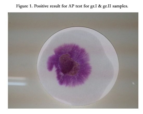
Figure 1. Positive result for AP test for gr.I & gr.II samples.
The second was the microscopic sperm identification test, it was done on all samples and the results were positive only in the samples categorized in group II due to the presence of sperms in the semen of fertile individuals (Figure 2), while results were negative in the samples categorized in group I due to the complete absence of sperms in the semen of Azoospermic individuals (Figure 3).
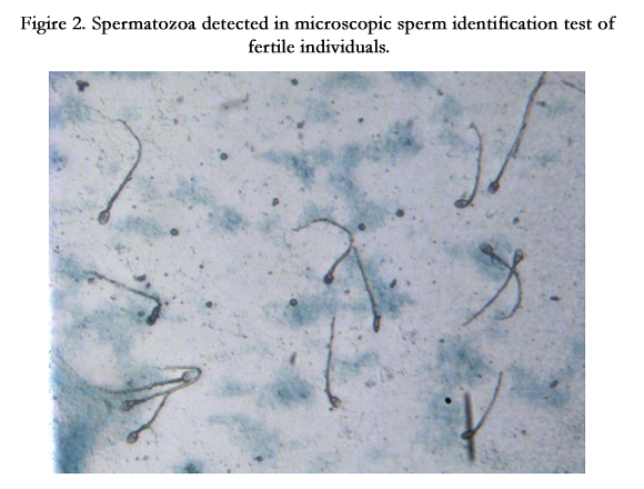
Figire 2. Spermatozoa detected in microscopic sperm identification test of fertile individuals.
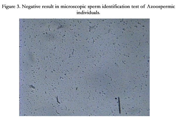
Figure 3. Negative result in microscopic sperm identification test of Azoospermic individuals.
DNA was extracted from all study samples (group I and group II), then quantitated using real-time PCR utilizing Quantifiler kit (Life Technologies). Table 1 shows the total genomic DNA concentrations for all samples in both groups.
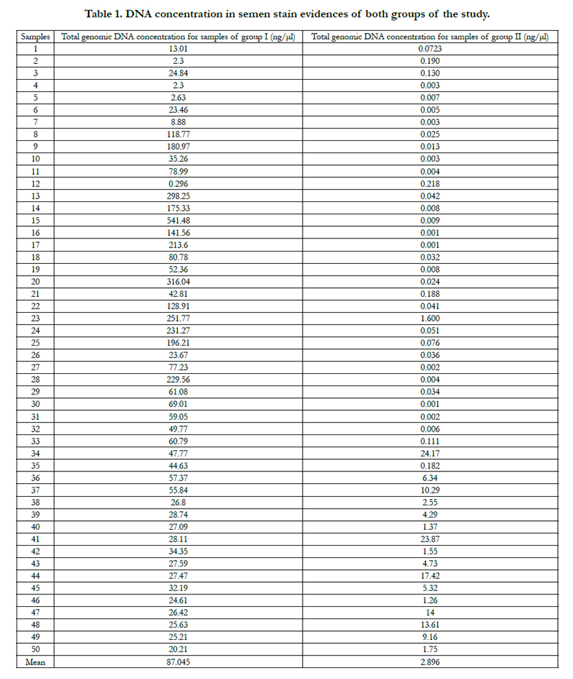

Table 1. DNA concentration in semen stain evidences of both groups of the study.
Multiplex fluorescent PCR was done for the 17 polymorphic YSTR loci included in AmpFLSTR® Yfiler® PCR Amplification Kit (Figure 4), then capillary electrophoresis for all amplified products was performed which showed complete STR profiles for all samples in both groups of the study.
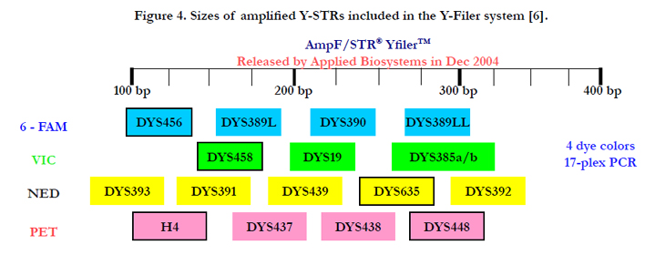
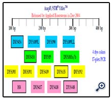
Figure 4. Sizes of amplified Y-STRs included in the Y-Filer system [6].
Discussion
In case of the occurance of such previous results of pre-DNA tests (acid phosphatase test & microscopic sperm identification test) in both groups of the study, the majority of criminal laboratories take the decision to do DNA profiling only for the samples of group I (fertile individuals) since it is confirmed that those types of suspected biological evidences are semen. Consequently, it is important to do DNA profiling for those samples in order to identify the man/men which is/are the source/sources of this semen. But, in case of investigating samples as those of group II (Azoospermic individuals), the decision would be not to continue investigating these samples as the criminal lab will not be assured that those suspected biological evidences are semen, for example; these samples can be vaginal secretions which gave the same positive results for presumptive acid phosphatase test and negative results for the confirmatory sperm identification test. Besides the fact that Azoospermic individuals are rarely present in the human population and accordingly it is very rare that the semen of one of them is that which is found, recovered and analyzed by the crime lab.
According to the results of real-time PCR shown in Table 1; by comparing the mean human DNA concentration for the samples of group I (87.045 ng/μl) with the mean human DNA concentration for the samples of group II (2.896 ng/μl), we notice a huge difference between them, which indicates that the DNA concentration in the semen of Azoospermic individuals reaches about 3.33% from the total DNA concentration in the semen of fertile individuals, which also means that spermatozoa constitute more than 96% from the total DNA sources constituted in the human semen. Other sources (≈ 4%) may include different types of cells.
Inspite of the detection of these minute amounts of DNA in the semen stain evidences of Azoospermic individuals, full Y-STR profiles were obtained in all samples categorized in group II. This revealed the success in the ability to extract and concentrate minute traces of genomic DNA present in the semen stains of Azoospermic individuals with an accuracy reaches 100%.
Conclusion
The methodology performed in this study to extract and concentrate trace amounts of DNA present in the semen stain samples of group II, has succeeded to obtain full Y-STR profiles from Azoospermic semen stain evidences. As a consequence, it is recommended to follow this methodology in case of performing DNA typing for suspected semen stain evidences recovered from crime scenes, to guarantee obtaining full DNA profiles even if those semen stain evidences were attributed to Azoospermic individuals.
Acknowledgement
This project was supported by the College of Forensic Sciences – Naif Arab University for Security Sciences (NAUSS).
References
- Gaensslen RE (1983) Sourcebook in Forensic Serology, Immunology, and Biochemistry, Research Foundation of the City University of New York: 101–114.
- Max M. Houck, Jay A. Siegel (2010) Fundamentals of Forensic Science (2nd edtn), Elsevier, The boulevard, Langford Lane Kidlington, Oxford, UK: 242-243.
- Qiagen (2012) QIAamp DNA Investigator Handbook (2nd edtn), Isolation of Total DNA from Body Fluid Stains: 20–22.
- Applied Biosystems. User’s Manual, Quantifiler® DNA Quantification Kit. Revised 3/12 USA (2012).
- Applied Biosystems. AmpFlSTR® Yfiler® PCR Amplification Kit User Guide. Applied Biosystems by Life Technologies Corporation (2012); 12–13, 21–22, 25–26, 57–60, 62–63.
- John M. Butler, Bruce R. McCord (2006) Y-STRs and mtDNA. (ppt). AAFS 2006 Workshop, Seattle, WA, February 20.
- Walter F Boron (2003) Medical Physiology: A Cellular and Molecular approach. Elsevier/Saunders: 1135-1136.
- Mann T (1954) The Biochemistry of Semen. London: Methuen & Co. New York: John Wiley & Sons. Retrieved November 9, 2013.
- Guyton, Arthur C (1991) Textbook of Medical Physiology (8th edtn) Philadelphia: W.B. Saunders: 890–891.
- Jarvi K, Lo K, Fischer A, Grantmyre J, Zini A, et al. (2010) CUA guideline: the workup of azoospermic males: Can Urol Assoc J; 4(3):163.
- Refaat AM (2012) Y-STR Profiling is a Unique and Valuable Tool in Forensic Investigation. International Journal of Medical Toxicology and Forensic Medicine; 2(4): 143-147.


