Effect of Some Antihypertensive Drugs on Alkaline Phosphatase and DNA of Mice
Ngabireng Marie. Claude1*, Menye Cyrille2, Kouam F.Simeon3, Ntede N .Hyppolite4, Tagoudjeu Jacques1, Awono Onana1
1.Chemistry Department, Faculty of Science, Mansoura University, Mansoura, Egypt.
2.Botany Department, Faculty of Science, Mansoura University, Mansoura, Egypt.
*Corresponding Author
OmAli Y. El-Khawaga,
Faculty of Community, King Khalid University, Rejal Almaa, Kingdom of Saudi Arabia.
E-mail: dr_elkhawaga@yahoo.com
Article Type: Research Article
Received: February 21, 2013; Accepted: March 15, 2013; Published: March 29, 2013
Citation: El-Khawaga OY, El-Waseef A, Y.O. Ellazec, M.M. El-Naggar, M. Abd alla (2013) Effect of Some Antihypertensive Drugs on Alkaline Phosphatase and DNA of Mice. Int J Clin Pharmacol Toxicol. 2(3), 58-62. doi: dx.doi.org/10.19070/2167-910X-1300012
Copyright: El-Khawaga OY© 2013. This is an open-access article distributed under the terms of the Creative Commons Attribution License, which permits unrestricted use, distribution and reproduction in any medium, provided the original author and source are credited.
Abstract
Various antihypertensive drugs are in a wide use for the treatment of hypertension. It is important to investigate the influences of chronic administration of these drugs on genomic DNA. So the objective of the present study was to elucidate the effects of chronic administration of some antihypertensive drugs e.g. amlodipine, atenolol and captopril on alkaline phosphatase(ALP) and genomic DNA in liver, spleen and kidney of Swiss albino mice. A total of 150 Swiss albino mice were divided into 10 groups (15 mice each): a control group and 3 groups to be treated by various doses of each of the three drugs. ALP was assayed in liver, spleen and kidney after drugs administration for 1; 3 and 6 months. The genotoxic effects of the three antihypertensive drugs were evaluated by RAPD-PCR technique. All studied drugs caused elevations in the activities of ALP in liver , spleen and kidney homogenates. Amlodipine induced DNA damage in the homogenates of liver, spleen and kidney . On the other hand, atenolol and captopril did not affect the genomic DNA. From our findings we can conclude that the chronic administration of the above antihypertensive drugs induce inflammation and amlodipine had toxic effect on the genomic DNA while atenolol and captopril had no apparent effects on DNA.
2.Materials and Methods
2.1.Chemicals
2.2.Experimental animals
2.3.Preparation of homogenates
2.4.Biochemical Analysis
2.5.Genomic DNA extraction and RAPD analysis
2.6.Statistical analysis
3.Results
4.Discussion
5.References
Introduction
Cardiovascular diseases are one of the leading causes of death for people in developed countries world-wide. Hypertension and osteoporosis are frequent dis -eases among elders. Both are induced by interaction of many genetic and environmental factors (Zhang et al., 2007). Many metabolic and physiological changes occur in hypertensive individuals and some of these processes stem from the associated increased oxidative stress ( Vasdev, 2006). The depression in antioxidant enzymes and increase in oxidants in the hypertensive state have been reported to increase the production of reactive oxygen species (Lassegue and Griendling, 2004 ; Manning et al., 2005 ). The reactive oxygen species/free radicals resulting from the oxidant-antioxi -dant imbalance tend to accumulate and are known to cause oxidative damage to the cellular macromolecules including the genetic material (Rao, 2009). The genetic instability can include mutations, chromosomal aber -rations and unscheduled DNA synthesis (Prerara and Bapat, 2007 ; Shimizu et al., 2008). Amlodipine is a derivative of dihydropyridine and is one of calcium chan -nel blockers. Its principal action is to inhibit calcium entry through voltage-gated transmembrane L-type channels, thus decreasing intercellular calcium concen -tration and inducing smooth muscle relaxation (Liu et al., 2007; Devabhaktuni and Bangalore, 2009; Liu el al., 2011). Atenolol is a beta 1-selective drug that binds to the beta-receptors and prevents the stimulation by cat -echolamine resulting in lowering of heart rate and of the systemic blood pressure (Wadworth et al., 1991). Captopril is an angiotensin-converting enzyme (ACE) inhibitor used for the treatment of hypertension and some types of congestive heart failure. Captopril was the first ACE inhibitor developed and was considered a break through both because of its novel mechanism of action and also because of the revolutionary devel -opment process (Akif et al., 2010).
Alkaline phosphatase (ALP) is an enzyme that cata -lyzes the hydrolysis of organic pyrophosphate and in -hibitor of vascular calcification (Harmey et al., 2004). Although ALP is expressed in a variety of tissues, its concentrations are highest in bone, liver and kidney (Schoppet and Shanahan, 2008). Accordingly, serum levels of ALP are used in clinical practice as a marker of hepatic , peripheral vascular and bony diseases (Regidor et al., 2008). No published data have been found in the relevant literature on the toxicity of anti -hypertensive drugs on the DNA of normal mice and thereby the present study was undertaken to determine the effect of chronic administration of recommended ; half recommended and one and half recommended doses of the antihypertensive drugs amlodipine, ateno -lol and captopril on the genomic DNA and on the ac -tivity of alkaline phosphatase (ALP) in the liver, spleen and kidney of Swiss albino mice.
Materials and Methods
Atenolol was purchased from Kahira Pharmaceutical. and Chemical Industries Company (Egypt). Amlodi -pine was purchased from Pfizer (Egypt) and captopril was purchased from Bristol Myers Squibb (Egypt). All other chemicals were of analytical grade.
All experiments were performed using adult female Swiss albino mice, with an average body weight of 25 g purchased from Theodore Bilharz Research Institute, Giza, Egypt. The mice were housed in steel mech cage and provided with commercial standard diet and tap water ad libitum.
A total of 150 Swiss albino mice were divided into 10 groups, 15 mice each, according to the following scheme: group 1, control (untreated) mice; groups 2-4, mice received captopril in doses of 1.3 mg/kg b.w. (low dose; half the recommended dose), 2.6 mg/kg b.w. (the recommended dose) and 3.9 mg/kg b.w. (high dose; one and half times the recommended dose) re -spectively every other day; groups 5 -7, mice received amlodipine in doses of 0.26 mg/kg b.w. (low dose; half the recommended dose), 0.52 mg/kg b.w. (the recom -mended dose) and 0.78 mg/kg b.w. (high dose; one and half times the recommended dose) respectively every other day and groups 8-10, mice received atenolol in doses of 5.2 mg/kg b.w. (low dose; half the recom -mended dose), 10.4 mg/kg b.w. (the recommended dose) and 15.6 mg/kg b.w. (high dose; one and half times the recommended dose) respectively every other day. Each of the nine groups of mice receiving the drugs were classified into subgroups, 5 animals each. The animals of the subgroups were sacrificed at 1 , 3, 6 months of starting administration of the drugs while the animals of the control group were all killed at the end of the experimental period. Liver, spleen and kidney were immediately excised, washed in ice-cold saline, blotted dry and weighed for measuring various biochemical parameters.
An accurately weighed piece of each of liver, spleen and kidney tissues was homogenized in ice-cold 0.9 % saline using a Teflon pestle connected to a homog-enizer motor. The homogenates were adjusted at a concentration of 5 %(w/v). The homogenates were centrifuged at 5000 rpm for 30 minutes at 4 0C to re -move cell debris and nuclei. The resulting supernatant was used for biochemical analysis.
Alkaline phosphatase activity was estimated by the method of Ali et al. (2006) using a commercial available assay kit (AAT Bioquest®, Inc., Egypt). Protein in the homogenates was determined by the method of Lowry et al.(1951).
Total DNA from mice tissues was extracted and purified according to the Wizard® Genomic DNA Purification Kit supplied by Promega Corporation (USA). Among several primers used for screening and RAPD-PCR analyses of the studied samples only one primer OPE-13: 5- - CCCGATTCGG-3- was chosen. Genomic DNA was analysised with random amplified polymorphic DNA polymerase chain reaction (RAPD-PCR) technique according to the method described by Williams et al. (1990). The PCR amplifications were performed in 25 µl of the reaction mixture contain -ing 1µl of template DNA, 3.5 µl primer and pure taq Ready- To- Go PCR beads. The PCR conditions were as follows: denaturing at 94 0C for 1.5 min, annealing at 37 0C for 1.5 min and extension at 72 0C for 3 min. The products were mixed with the loading buffer and were loaded in 2% agarose gels and electrophoresed at 100 V for 1h. The gels were stained with ethidium bromide and visualized under UV light.
The results are expressed as means ± SD. Statistical analysis was performed according to the method of Murray (1982). Data were analyzed using unpaired Student,s t-test. P values of < 0.05 were considered to be statistically significant.
Results
Table 1 illustrates the alkaline phosphatase (ALP) ac -tivities in mice of groups. After administration of captopril for one , three and six months in doses of 1.3, 2.6 , 3.6 mg/kg b.w., highly significant elevations in the activities of ALP in the liver, spleen and kidney ho -mogenates were found compared to its activities in the homogenates of the control group. Similarly, a highly significant increases in the activities of ALP in the liver and spleen homogenates of mice were observed after administration of 5.2, 10.4, 15.6 mg/kg b.w. atenolol for one, three and six months. On the other hand, a significant decrease was observed in ALP activity in the kidney homogenates after administration of 5.2 mg/kg b.w. atenolol for one month while no significant changes were found after three and six months from the administration of atenolol. Moreover, a significant decrease in the kidney ALP activity on the administra -tion of 10.4 and 15.6 mg/kg b.w. of atenolol for six months was found compared to the control activity. The administration of 0.26 , 0.52 and 0.78 mg/kg b.w. of amlodipine for one, three and six months caused highly significant increases in the activities of ALP in the liver, spleen and kidney homogenates compared to the corresponding activities of the controls.
The results of RAPD-PCR for DNA samples isolated from liver, kidney and spleen of control mice and mice treated for 1, 3 and 6 months with various doses of captopril , atenolol or amlodipine are shown in Fig 1A, B, C. The amplification of products obtained by this technique showed the presence of numerous bands between 2 and 23 kbp (Fig 1C). The primer gave a recognizable profile of bands for amlodipine treated samples (Fig 1C) for 6 months compared to atenolol, captopril (Fig 1 A & B) and control samples (Fig 1 A & C). Overall, the treatment by amlodipine caused more changes in RAPD-PCR patterns than treatment by either captopril or atenolol especially in liver, spleen and kidney.
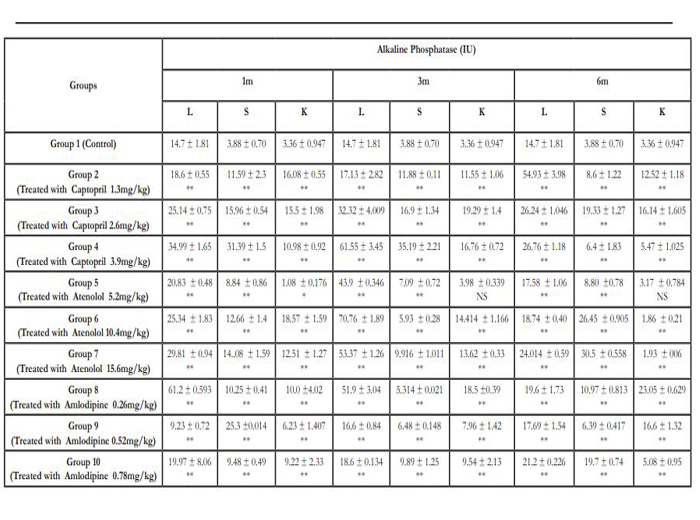

Table 1: Alkaline phosphatase in liver (L) , spleen (S) and kidney (K) of control mice and mice treated with various doses of Captopril, Atenolol and Amlodipine.
**highly significant compared to the corresponding control level.
1 m, 3m and 6 m: samples after treatment for 1, 3 and 6 months respectively.
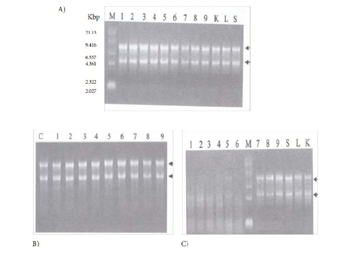
Figure 1: RAPD- PCR analysis of DNA samples isolated from livers (lanes 1-3); spleen (lanes 4-6) and kidney (lanes 7-9) of capotpril –treated (A) ; atenolol- treated (B) and amlodipine-treated mice (C) : Lane M :standard molecular weight marker . Lanes K, L , S in Fig. A and C: kidney, liver and spleen DNA of control mice. Lane C in Fig. B: liver DNA of control mice. Lanes 1, 4,7 : samples after one month treatment ; Lanes 2, 5, 8: samples after 3 month treatment; Lanes 3, 6, 9: samples after 6 month treatment.
Discussion
The hypertension increases the risk for stroke and coronary heart disease and is a main contributor to pre-mature death (Yadav et al., 2008). Toxic effects of drugs may be functional, biochemical, structural and specific. Such effects are evaluated by assessment of oxidant-antioxidant parameters in tissues and severity of DNA damage (Polat et al., 2010). The pathology of essential hypertension may involve one or more abnormalities in cardiovascular homoeostatic mecha -nisms including endothelial dysfunction (Beevers et al., 2001). The endothelial dysfunction is related to a de -fect in vasodilator nitric oxide (NO) synthesis. There may be an elevation of reactive oxygen species like superoxide anion, which inactivate the NO. Normally, the endogenous antioxidant mechanism involving en -zymes like catalase and superoxide dismutase in addi -tion to reduced glutathione scavenge and regulate the superoxide formed. This protective mechanism may be defective in essential hypertension cases (Touyz, 2004). In the present study the effects of treatment with the antihypertensive drugs amlodipine, atenolol and captopril on alkaline phosphatase (ALP) activity and genomic DNA was investigated in liver, spleen and kidney of mice. After chronic administration of captopril and amlodipine, the activity of ALP in liver , spleen and kidney homogenates were highly signifi -cantly increased compared to its level in the organs of mice. These finding are in accordance with those re-ported by Nishiya and Sugimoto, (2001) , Gradosova et al. (2011) and Moraes et al. (2011). Tonelli et al. (2009) reported that the higher level of ALP can be used as a marker for patients with an underlying inflammatory state, vascular calcification and as a liver function. Some investigators showed that the chronic administration of captopril affected the promotion of the secretion of ALP and its level depends on the dose of captopril (Liu et al. 2011). The present results demonstrated that chronic administration of atenolol exhibited signifi -cant increase in the activity of ALP. These results are in good agreement with those of other investigators (Mota et al., 2008). The elevation of ALP activity in normal mice after administration of antihypertensive drugs may reflect a link between the abnormal mineral metabolism of kidney and excess cardiovascular mor-tality (Ali et al., 2006 & Blayney, et al., 2008).
The analysis of genomic alterations in liver, spleen and kidney of mice treated with atenolol and capto -pril using RAPD-PCR fingerprinting showed that these drugs did not produce significant changes in the RAPD-PCR fingerprints of the liver, spleen and kid -ney genomes. Both drugs produced nearly the same DNA banding patterns similar to that of control (non-treated) mice. However, amlodipine exerted an ad -verse affect on the DNA content of the liver, spleen and kidney. Apparently, amlodipine degraded the liver, spleen and kidney DNA and masked the sites for the annealing of the RAPD primers. These present finding are disagreeable with previous studies reporting that long term therapy of atenolol increased expression of fragile sites of DNA and caused chromosomal aber -rations (Telez et al., 2000 ; Telez et al., 2010). On the other hand, the present results are in consistent with the results of other investigators (Kang et al., 1997 & Liu et al., 2006) who reported that atenolol and capto -pril did not show any significant antiproliferative effect on the cultured rat aortic smooth muscle cells through -out any of the studied doses. Many studies indicated that the calcium channel blocker amlodipine influences DNA and affect bone tissues resulting in decrease in bone resorption (Rejnmork et al., 2006 ; Ushijima et al., 2010 ; Subash et al., 2010). In these previous re -ports, accumulating data suggested that this calcium channel blocker (amlodipine) has antiinflammatory-antioxidant and antiapoptotic properties beside its an -tihypertensive action (Yamagata et al., 2004 & Yoshii et al., 2006). To summarize, our results clearly indicated that the chronic administration of amlodipine affected the genomic DNA and the activity of ALP. The pre -sent results may be attributed to: [1] the balance be -tween inorganic pyrophosphate and serum phosphate which regulates vascular calcification. [2] imbalance in oxidant and antioxidant levels due to hypertension which resulted in accumulation of DNA damage with time ( Khanna et al., 2008). In addition it is concluded that the studied antihypertensive drugs may induce in -flammation by increasing the activity of ALP.
References
- Akif, M.; Georgiadis, D.; Mahajan, A.; Dive, V.; Sturrock, E. D.; Isaac, R. E.; Acharya, K. R. (2010). “High-Resolution Crystal Struc-tures of Drosophila melanogaster Angiotensin-Converting Enzyme in Complex with Novel Inhibitors and Antihypertensive Drugs”. Journal of Molecular Biology 400 (3): 502–517.
- Ali AT, Penny CB, Paiker JE, Psaras G, Ikram F, Crowther NJ. (2006) The effect of alkaline phosphatase inhibitors on intracellular lipid accumulation in preadipocytes isolated from human mammary tissue. Ann Clin Biochem, 43, 207 – 13.
- Beevers, G., Lip, G.Y.H. and O, Brien ,E. (2001): ABC of hy -pertension. The pathophysiology of hypertension. B.M.J. 322:912-916.
- Blayney, M.J.; Pisoni, R.L.; Bragg-Gresham J.L.; Bommer, J.; Piera, L. and Saito, A. (2008): High alkaline phosphatase levels in hemo -dialysis patients are associated with higher risk of hospitalization and death. Kidney Int, 74: 655- 663.
- Devabhaktuni, M. and Bangalore, S.(2009): Fixed combination of amlodipine and atorvastatin in cardiovascular risk management. Vasc Health Risk Manag, 5: 377-387.
- Gradosova, I.; Zivna, H.; Svejkovska, K.; Palicka, V.; Tichy, A. and Zivny, P.(2011): Effects of amlodipine on bone metabolism in male albino Wistar rats. Actavet. Brno, 80: 391-396.
- Harmey, D.; Hessle, L.; Narisawa, S.; Johnson, K.A.; Ter-keltaub, R. and Millan, J.L. (2004): Concerted regulation of inorganic pyrophosphate and osteopontin by alp2, enpp, and ank: an integrated model of the pathogenesis of mineralization disorders. Am J Pathol, 164: 1199- 1209.
- Khanna, H.D.; Sinha, M.K.; Khanna, S. and Tondon, R. (2008): Oxidative stress in hypertension: Association with antihypertensive treat-ment. Indian J Pharmacol. 52(3): 283-287.
- Lassegue, B., Griendling, K.K.; (2004): Reactive oxygen spe -cies in hypertension: an update. Am. J. Hypertens. 17:852-860.
- Liu, LL.; Li, QX.; Xia, L. Li, J. (2007): Differential effects of dihydropyridine calcium antagonists on doxorubicin- induced nephro -toxicity in rats. Toxicology, 231: 8190.
- Liu, YY; Cui, L and Wu, T. (2006): Effects of captopril on os -teoblast isolated from neonatal rat calvarium in vitro. Chinese J of Ger -ontology, 2006-02
- Liu, YY; Yao, WM; Wu, T; Xu BL; Chen, F. and Cui, L.(2011): Captopril improves osteopenia in ovariectomized rats and promotes bone formation in osteoblasts. J bone Miner Metab. 29(2): 149-58.
- Lowry, O. H.; Rosebrough, N. J.; Farr, A. and Randall, R. J. (1951) Protein measurement with folin-phenol reagent. J. boil. Chem., 193, 265- 275.
- Manning, RD,; Jr., Tian, N., Meng, S. (2005): oxidative stress and antioxidant treatment in hypertension and associated renal damage. Am. J. Nephrol, 25:311-317.
- Moraes, RB.; Correa, L. and Luz, JG. (2011): Adverse effects of the amlodipine on bone healing of the mandibular fracture: an experi -mental study in rats. Oral Maxillofac Sur. 15(2):93-101.
- Mota, A.; Silva, P.; Neves, D.; Lemos, C. and Calhau, C. (2008): Characterization of rat heart alkaline phosphatase isoenzymes and mod -ulation of activity. Braz J Med Biol Res, 41(7): 600-9.
- Murry, R.S.(1982): Schaum,s Outline Series of Theory and Problems of Probability and Statistics, Singapore, McGraw-Hill Book Company, vol, 8: pp265-298.
- Nishiya, Y and Sugimoto, S. (2001). Effects of various an-tihypertensive drugs on the function of osteoblast. Biol Pharm Bull, 24(6):628-33.
- Perera, S.; Bapat, B.(2007): Genetic instability in cancer. Atlas of genetics and cytogenetics in oncology and haematology. http://At-lasGeneticsOncology.org/Deep/GenetInstabilityCancerID20056.html
- Polat, B.; Suleyman, H.; Alp, H.H. (2010): Adaptation of rat gastric tissue against indomethacin toxicity. Chem Biol Interact, 186(1): 82-89.
- Rao, KS. (2009): Free radical induced oxidative damage to DNA: Relation to brain aging and neurological disorders. Indian Journal of Biochemistry and Biophysics, 46: 9-15.
- Regidor, D.L.; Kovesdy, C.P.; Methrotra, R.; Rambod, M.; Jing, J.; McAllister, C.J. and van Wyck, D. (2008): Serum alkaline phosphatase predicts mortality among maintenance hemodialysis patients. J Am Soc Nephrol, 19:2193-2203.
- Rejnmark, L.; Vestergaard, P. and Mosekilde, L.(2006): Treat -ment with beta- blockers. ACE inhibitors and calcium-channel blockers is associated with a reduced fracture risk: a nationwide case- control study. J Hypertens. 24: 581-589.
- Schoppet, M. and Shanahan, C.M. (2008): Role for alkaline phosphatase as an inducer of vascular calcification in renal failure. Kid -ney Int, 73: 989- 991.
- Shimizu, H.; Nakagami, H.; Osako, MK.; Hanayama, R.; Kunugiza, Y.; and Kizawa, T.(2008): Angiotensin II accelerates osteopo-rosis by activating osteoclasts. FASEB J 22:2465-2475.
- Subash, P.; Premagurumurth, K.; Sarasabharathi, A. and Che -rian, K.M. (2010): Total antioxidant status and oxidative DNA damage in a South Indian population of essential hypertensives. J of human Hyper -tension, 24: 475- 482.
- Telez, M.; Martinez, B.; Criado, B.; Lostao, C.M.; Penagarikano, O.and Ortega, B. (2000): In vitro and in vivo evaluation of the antihy-pertensive drug atenolol in cultured human lymphocytes: effects of long-term therapy. Mutagenesis, 15(3): 195-202.
- Telez, M.; Eduardo, OL.; Gonzalezc AJ.; Floresd, P. (2010): Assessment of the genotoxicity of atenolol in human peripheral blood lymphocytes: Correlation between chromosomal fragility and content of micronuclei. Mutation Research. 695: 46-54.
- Tonelli, M.; Curhan, G.; Pfeffer, M.; Sacks, F. and Thadhani, R. (2009): Epidemiology and prevention relation between alkaline phos -phatase, serum phosphate and all- cause or cardiovascular mortality. Cir -culation, 120:1784- 1792.
- Touyz, R.M.( 2004): Reactive oxygen species, vascular oxida -tive stress and redox signaling in hypertension, what is the clinical signifi -cance? Hypertension, 44: 248-252.
- Ushijima, K.; Liu, Y.; Maekawa, T.; Ishikawa, E.; Motosugi, Y. and Ando, H. (2010): Protective effect of amlodipine against osteoporo -sis in stroke- prone spontaneously hypertensive rats. Eur. J Pharmacol. 635: 227-230.
- Vasdev, S.; Gill, VD.; and Singa PK. (2006): Modulation of oxidative stress-induced changes in hypertension and atherosclerosis by antioxidants. Exp Clin Cardiol, 11(3):206-216.
- Wadworth, A. N.; Murdoch, D.; and Brogden, R.N. (1991): At-enolol, a reappraisal of its pharmacological properties and therapeutic use in cardiovascular disorders. Drugs, 42(3): 468- 510.
- Williams, J.G.K.; Kubellik, A.R.; Livak, K.J.; Rafalski, J.A. and Tingy, S.V. (1990): DNA polymorphism amplified by arbitrary primers are useful as genetic markers. Nucleic Acid Research, 18(22): 6531-6535.
- Yadav, S.; Boddula, R.; Genitta, G.; Bhatia, V. and Bansal, B. (2008): Prevalence and risk factors of pre-hypertension and hypertension in an affluent north Indian population. Indian J Med Res, 128: 712-720.
- Yamagata, K.; Ichinose, S. and Tagami, M. (2004): Amlodi -pine and carvedilol prevent cytotoxicity in cortical neurons isolated from stroke-prone spontaneously hypertensive rats. Hypertens Res, 27: 271-282.
- Yoshii, Y.; Iwai, M. and Li, Z. (2006): Regression of atheroscle -rosis by amlodipine via antiinflammatory and antioxidative stress actions. Hypertens Res, 29: 457- 466.
- Zhang X.; Sun Z.; Zheng L.; Li, Z; Liu, S.; Xu C. (2007): Preva-lence of dyslipidemia and associated factors among the hypertensive ru -ral Chinese population. Archives of Medical Research, 38(4): 432-439.


