Kissing Canines Associated with a Dentigerous Cyst: Case Report and Mini Review
Omami G1*, Mathew R2
1 Assistant Professor, Department of Oral Health Practice, University of Kentucky College of Dentistry, USA.
2 Assistant Professor, Department of Oral and Maxillofacial Radiology, Midwestern University College of Dental Medicine, USA.
*Corresponding Author
Galal Omami, BDS, MSc, MDentSc,
Diplomate, American Board of Oral and Maxillofacial Radiology,
Assistant Professor, Department of Oral Health Practice, University of Kentucky College of Dentistry,
800 Rose Street, Lexington, KY 40536,
USA.
Tel: (859)323 9833
Fax: (859) 323-9136
E-mail: jellodent@yahoo.com
Article Type: Case Report
Received: April 29, 2015; Accepted: May 29, 2015; Published: June 03, 2015
Citation: Omami G, Mathew R (2015) Kissing Canines Associated with a Dentigerous Cyst: Case Report and Mini Review. Int J Dentistry Oral Sci. 2(6), 84-86. doi: dx.doi.org/10.19070/2377-8075-1500018
Copyright: Omami G© 2015. This is an open-access article distributed under the terms of the Creative Commons Attribution License, which permits unrestricted use, distribution and reproduction in any medium, provided the original author and source are credited.
Abstract
Dentigerous cyst is the most common developmental odontogenic cyst of the jaws that encloses the crown of an unerupted tooth; however, involvement of more than one unerupted tooth in a single dentigerous cyst is extremely rare. In this report, we present a rare case of a single dentigerous cyst associated with the imapcted mandibular permanent canines. Literature is briefly reviewed and etiopathogenesisis hypothesized.
2.Introduction
3.Case Report
4.Discussion
5.Conclusion
6.References
Keywords
Dentigerous Cyst; Unerupted; Impacted; Cone Beam CT.
Introduction
Dentigerous cyst is the most common developmental odontogenic cyst and is associated with the crown of an unerupted tooth and attached to its cervix. However, dentigerous cyst may be seen in association with impacted supernumerary tooth or odontoma. It likely arises by the accumulation of fluid between reduced enamel epithelium and the tooth crown. The most common site is mandibular third molar region followed by maxillary permanent canine, mandibular premolars, and maxillary third molars. Dentigerous cysts are often asymptomatic and are incidentally discovered during routine radiographic examination; however, they can cause jaw expansion, cortical erosion, and root resorption. Our literature review showed only a handful of cases of a single dentigerous cyst involving multiple permanent teeth. In the present report, we present a rare case of a single dentigerous cyst associated with the imapcted mandibular permanent canines.
Case Report
A 12-year-old Caucasian girl was presented to the department of orthodontics for evaluation of dentition. Patient’s medical history was unremarkable. On clinical examination, there were multiple retained deciduous and missing permanent teeth. Cone beam CT (CBCT) scan has shown multiple congenitally missing and unerupted permanent teeth. Lower permanent canines were horizontally and deeply impacted with their crowns located just inferior and facial to the apices of lower incisors. Both crowns were surrounded by a well-defined round corticated radiolucency that has caused slight thinning and expansion of the facial cortex of the mandibular symphysis. The overall estimated measurements of the lesion were 14.2 x 8.2mm in maximum vertical and horizontal dimensions, respectively. The lesion appeared to be attached to the cement-enamel junctions (CEJs) of both teeth. No root resorption or displacement of lower incisors has been appreciated. A provisional diagnosis of dentigerous cyst was suggested. However, the expansile behavior of the lesion ruled out the possibility of hypertrophic follicle. Surgical management included enucleation of the lesion along with removal of the associated impacted canines. The specimen was sent for histopathological examination that confirmed the diagnosis of dentigerous cyst.
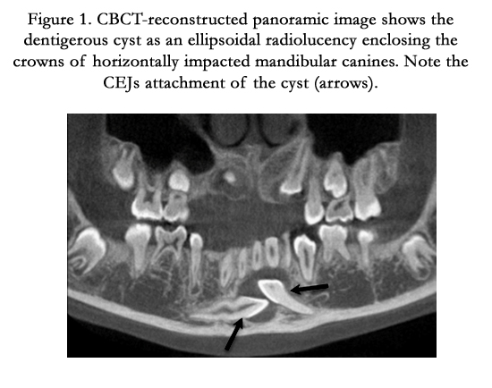
Figure 1. CBCT-reconstructed panoramic image shows the dentigerous cyst as an ellipsoidal radiolucency enclosing the crowns of horizontally impacted mandibular canines. Note the CEJs attachment of the cyst (arrows).
Discussion
Dentigerous cyst is the most common developmental odontogenic cyst of the jaws that encloses the crown of an unerupted tooth by expansion of its follicle, and is attached to its cervix. It has previously been known as follicular cyst; however, Browne et al. stressed that the term “dentigerous” (tooth-bearing) is preferable to that of “follicular cyst”, as the latter implies a derivation from the tooth follicle which is a mesodermal structure [1]. Another reason for not adopting the term “follicular cyst” is that this is most commonly used to refer to follicular cysts of the ovary and hair follicle as well. Some unerupted teeth have a slightly enlarged follicle; this does not necessarily signify a cyst, or even a potential cyst unless the pericoronal width is at least 5mm [2]. However, it is hardly possible to distinguish between a large dental follicle and a small dentigerous cyst [2]. Mourshed has calculated that the frequency of dentigerous cysts is 1.44 for every 100 unerupted teeth [3]. Three radiological variations of dentigerous cyst may be observed. The central variant where the crown is enveloped symmetrically, the lateral type in which the cyst appears on one aspect of the crown, and the circumferential variant as the entire tooth appears to be enveloped by cyst.
It has been suggested that dentigerous cysts may develop by accumulation of fluid either between the reduced enamel epithelium and the enamel or between layers of reduced enamel epithelium. Main has proposed that the pressure exerted by a potentially erupting tooth on an impacted follicle results in obstruction of the venous outflow and thereby induces rapid transudation of serum across the capillary walls, and the increased hydrostatic pressure of this pooling fluid in turn separates the follicle from the crown [4]. Toller believed that the likely origin of dentigerous cyst was a breakdown of proliferating cells of the follicle following impeded eruption that contributes to the increase in intrafollicular osmotic tension and subsequently to further expansion of the follicle [5].
Kissing teeth are unusual radiographic findings. The term was first described by Van Hoof in 1973 when he reported a case of impacted permanent molars that have occlusal surfaces contacting each other within a single follicular space with roots of each pointing in opposite directions [6]. Kissing molars are sparsely reported in literature. To best of our knowledge, this is the first case to describe “kissing canines”.
Involvement of dentigerous cyst with more than one unerupted tooth is exceedingly rare. To best of our knowledge, only a few cases of dentigerous cysts associated with multiple unerupted teeth have been reported in English literature [7-12] (Table 1). In the present case, the association of the impacted mandibular canines was unequivocally evident on basis of imaging and surgical grounds. However, the exact follicle of origin was not clearly identified. So it was not clear if coalescence has occurred between two adjacent dentigerous cyst linings or between the lining of a pre existing cyst and the reduced enamel epithelium of an adjacent follicle. Another remote possibility is that the tooth tends to erupt in the cyst lining in the same fashion as it does in the oral mucosa [2]. This hypothesis does not seem to hold true at least in case of dentigerous cyst where only the crown is surrounded by the lesion.
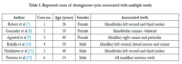

Table 1. Reported cases of dentigerous cysts associated with multiple teeth.
Conclusion
In conclusion, in this report we present a new radiographic, if not pathologic, variant of dentigerous cyst. We assume that meticulous radiographic analysis of dentigerous cysts could be helpful in speculating thoughts on etiology.
References
- Browne RM, Smith AJ (1991) Pathogenesis of odontogenic cysts: In Investigative Pathology of the Odontogenic Cyst, Browne RM. (1st edn), New Jersey, CRC Press Boca Raton. 88–109.
- Shear M (1994) Developmental odontogenic cysts. An update. J Oral Pathol Med 23(1): 1-11.
- Mourshed F (1964) Aroentgenographic study of dentigerous cysts: incidence in a population sample. Oral Surg Oral Med Oral Pathol 18: 47-53.
- Main D (1970) The enlargement of epithelial cyst of jaws. Odontologisk Revy 21: 29- 49.
- Toller P (1966) Epithelial discontinuity in cyst of the jaws. Br Dent J 120(2): 74-78
- Van Hoof RF (1973) Four kissing molars. Oral Surg Oral Med Oral Pathol 35(2): 284.
- Robert H Ivy (1939) Multiple dentigerous cysts with special reference to occurrence in siblings. Annals Surg 109(1): 114-125.
- Gonzalez MS, Spalding MP, Payne BJ, Giannini JP (2011) Adentigerous cyst associated with bilaterally impacted mandibular canines in a girl: a case report. J Med Case Rep 5: 230.
- Agrawal M, Raghavendra P, Singh B, Agrawal N (2011) Multiple teeth in single dentigerous cyst follicle: A perplexity. Ann MaxillofacSurg 1(2): 187- 189.
- Rohilla M, Namdev R, Dutta S (2011) Dentigerous cyst involving multiple impacted teeth. A rare case report. J Indian Soc Pedod Prev Dent 29: 244-247.
- Nishikawa S, Cheng J, Koyano Y, Nomura T, Kato T, et al. (1996) Dentigerous cyst involving two impacted molars: report of a case. Oral Med Pathol 1(1): 60-62.
- Ganesh P, Anehosur V, Joshi A, Gopalkrishnan K (2012) Dentigerous cyst of maxilla involving multiple impacted teeth: a rare case report. Int Joral Maxillofac Path 3(3): 45-49.


