Langerhans Cell Histiocytosis: Letterer - Siwe Disease Interest of Dermatological Signs for Diagnosis
Elmahi H*, Lahlou A, Gallouj S, Mernissi FZ
Department of Dermatology, University Hospital Hassan II, Fez, Morocco.
*Corresponding Author
Hakima Elmahi,
Department of Dermatology,
University Hospital Hassan II, Fez, Morocco.
E-mail: elmahi.hakima@gmail.com
Received: February 16, 2017; Accepted : May 09, 2017; Published: May 18, 2017
Citation: Elmahi H, Lahlou A, Gallouj S, Mernissi FZ (2017) Langerhans Cell Histiocytosis: Letterer - Siwe Disease Interest of Dermatological Signs for Diagnosis. Int J Clin Dermatol Res. 5(4), 114-115. doi: http://dx.doi.org/10.19070/2332-2977-1700030
Copyright: Elmahi H© 2017. This is an open-access article distributed under the terms of the Creative Commons Attribution License, which permits unrestricted use, distribution and reproduction in any medium, provided the original author and source are credited.
Abstract
Langerhans cell histiocytosis is defined as a clonal proliferation of Langerhans phenotypic-like cells. Letterer-Siwe disease is the most common and serious of these entities, affecting mainly infants up to two years of age. We present a case of this rare disease, diagnosed after dermatological examination, highlighting its typical aspects.
2.Introduction
3.Case Report
4.Discussion
5.Conclusion
6.References
Keywords
Langerhans Cell Histiocytosis; Infant; Letterer - Siwe Disease.
Introduction
Langerhans cell histiocytosis (LCH) is a rare disorder that involves the clonal proliferation of the Langerhans cells. LCH is often diagnosed in childhood, but any age group can be affected, from infancy through adulthood [1]. The cause of this disease is unknown, although many possibilities have been explored. The clinical presentation is highly variable. The severity and prognosis depend on the type and extent of organ involvement [3]. The disease classically presents with three syndromes namely Eosinophilic granuloma, Letterer-Siwe disease and Hand-Schuller-Christian disease [4]. Letterer-Siwe disease is the most common and serious of these entities, affecting mainly infants up to two years of age [5]. We present a case of this rare disease, diagnosed after dermatological examination, highlighting its typical aspects.
Case Report
A 5-month-old female infant presented with a progressively worsening generalized rash of 1 month duration, fever, poor feeding, and abdominal distension. The examination showed jaundice, widespread petechias, telangiectasias, small erythematous papules, ulcerative - necrotic lesion and erythematous - vesical - crustry - flaky - purpuric lesions on the scalp, trunk, extremities of upper limbs (especially palmar region) and diaper area [Figure 1 and 2]. There was also a necrotic lesion on the palate and purpuric - hemorrhagic lesions on the oral cavity [Figure 3]. An initial laboratory workup revealed anemia, thrombocytopenia, and hepatosplenomegaly. A working diagnosis of Langerhans cell histiocytosis was reached that was later confirmed with a skin biopsy. Two day later, the patient evolved to death due to multiple organs failure.
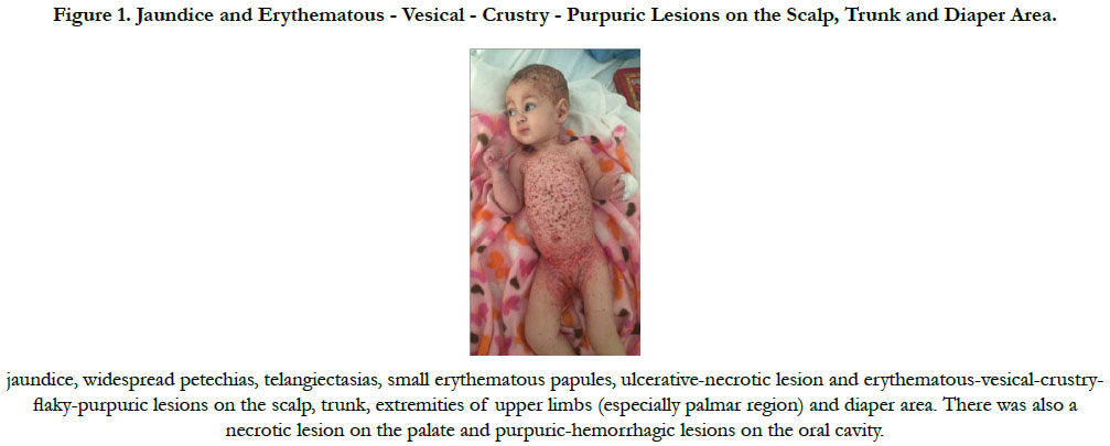
Figure 1. Jaundice and Erythematous - Vesical - Crustry - Purpuric Lesions on the Scalp, Trunk and Diaper Area.
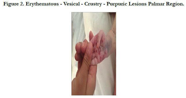
Figure 2. Erythematous - Vesical - Crustry - Purpuric Lesions Palmar Region.
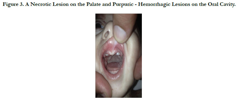
Figure 3. A Necrotic Lesion on the Palate and Purpuric - Hemorrhagic Lesions on the Oral Cavity.
Discussion
Letterer - Siwe disease (LSD) is a kind of a histiocytosis, rarely seen in clinic. However, its prognosis is poor. Children with less than two years of age, with involvement of the hematopoietic system, skin, liver, lungs, and spleen, even with aggressive treatment, may have mortality greater than 50% [3, 5]. The differential diagnosis is extensive and includes seborrheic dermatitis, eczema, diaper dermatitis, Darier’s disease, urticaria pigmentosa, arthropod bites, scabies, leukemia, B- and T-cell lymphomas, and some of the non-Langerhans cell histiocytoses (especially indeterminate cell histiocytosis, benign cephalic histiocytosis, generalized eruptive histiocytosis, and xanthoma disseminatum). Ear involvement can easily be mistaken for chronic otitis media or otitis externa [6]. The diagnosis of LCH is based on morphological, immunohistochemical and ultrastructural features. The standard therapy length recommended for LCH involving the spleen, liver, or bone marrow (high-risk organs) is now 12 months based upon the DALHX- 83 and HISTSOC-LCH-III (NCT00276757) studies. In the Histiocyte Society LCH-II and LCH-III studies, the standard arm consisted of vinblastine and prednisone as described above under multifocal bone, but 6-mercaptopurine was added to the continuation phase of the protocol [7].
Conclusion
To conclude, the LSD is a rare condition, but, due to its severity, dermatologists should be able to recognize its typical dermatological signs, which may be decisive to define the treatment and prognosis of affected patients.
References
- Narges Gholami (2016) Pericardial Effusion in Langerhans Cell Histiocytosis: A Case Report. Iran Red Crescent Med J. 18(6): e25604.
- Mehes G, Irsai G, Bedekovics J, Beke L, Fazakas F, et al., (2014) Activating BRAF V600E mutation in aggressive pediatric Langerhans cell histiocytosis: demonstration by allele-specific PCR/direct sequencing and immunohistochemistry. Am J Surg Pathol. 38(12): 1644-8.
- Agarwal P, Kaushal M (2014) An unusual presentation of Langerhans cell histiocytosis. J Cytol. 31(4): 227–9.
- Azariah ED, Chandrasekaran D, Chinnaswami R, Balasubramaniam S, Jagdish E (2016) "Histiocytosis X" - A Rare Case Report. J Clin Diagn Res. 10(10): ZD19-ZD22.
- Ferreira LM, Emerich PS, Diniz LM, Lage L, Redighieri I (2009) Langerhans cell histiocytosis: Letterer-Siwe disease - the importance of dermatological diagnosis in two cases. An Bras Dermatol. 84(4): 405-9.
- Jeffrey P Callen, Joseph L Jorizzo, John J Zone, Warren W Piette, Misha A Rosenbach, et al., (2017) Dermatological Signs of Systemic Disease. Pages 183-191.
- National Cancer Institute (2016) Langerhans Cell Histiocytosis Treatment (PDQ®): Health Professional Version. PDQ Pediatric Treatment Editorial Board. Bethesda: US.


