Sporotrichoid - Like Tuberculosis in a Child: Unusual Clinico - Dermoscopic Aspect
C. Saadani Hassani*, H. Baybay, J. Ziani, M. Rimani, F.Z. Mernissi
Departement of Dermatology, CHU Hassan II, Fez, Morocco.
Laboratoty Hassan, Rabat, Morocco.
*Corresponding Author
C.Saadani Hassani,
Departement of Dermatology, CHU Hassan II, Fez, Morocco.
E-mail: ch.saadani@gmail.com
Received: January 26, 2019; Accepted : October 21, 2019; Published: October 22, 2019
Citation: C. Saadani Hassani, H. Baybay, J. Ziani, M. Rimani, F.Z. Mernissi. Sporotrichoid - Like Tuberculosis in a Child: Unusual Clinico - Dermoscopic Aspect. Int J Clin Dermatol Res. 2019;7(5):241-242. doi: dx.doi.org/10.19070/2332-2977-1900053
Copyright: C. Saadani Hassani© 2019. This is an open-access article distributed under the terms of the Creative Commons Attribution License, which permits unrestricted use, distribution and reproduction in any medium, provided the original author and source are credited.
Cutaneous tuberculosis simulating sporotrichosis has been sporadically reported.; it mainly affects children after post-traumatic inoculation [1]. We describe a new pediatric case of gummy tuberculosis with a sporotrichoid pattern, as well as its dermoscopic appearance.
A child of 10 years who has been vaccined by BCG just 1 week after born, presented since 4 months, with two lesions on his right arm. On examination, it was the primary nodule formed two erythematous nodules developed subsequently over his right arm in a linear distribution, simulating sporotrichosis. The oldest was ulcerated at its center, well-defined, with a regular accentuated borders (Figure 1a). Dermoscopically, the lesion in exhibits a linear and hairpin vessels, and whitish reticular streaks at the periphery (Figure 2a). The recent plaque was erythemato-squamous, well limited (Figure 1a), with linear and hairpin vessels on dermoscopy (Figure 2b). Unilateral axillary lymph nodes were involved with a 3cm, firm and mobile lymphadenopathy. A skin and lymph node biopsy were performed, The histopathology of the primary nodule disclosed dermal epithelioid cell granuloma with caseation necrosis and peripheral lymphocytic infiltration (Figure 3). Ziehl-Nielsen, periodic acid-Schiff or Grocott’sstaining did not provided any additional positive information.The primary clinical differential diagnosis included sporotrichosis and leishamiosis sporotrichosis- like. We started anti-tuberculosis drugs for 6 months. After the initiation of the therapy his cutaneous lesions rapidly subsided with some residual scar formation (Figure 4a, b).
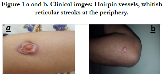
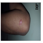
Figure 1 a and b. Clinical imges: Hairpin vessels, whitish reticular streaks at the periphery.
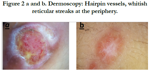
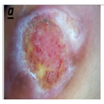
Figure 2 a and b. Dermoscopy: Hairpin vessels, whitish reticular streaks at the periphery.
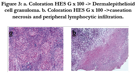
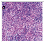
Figure 3: a. Coloration HES G x 100 -> Dermalepithelioid cell granuloma. b. Coloration HES G x 100 ->caseation necrosis and peripheral lymphocytic infiltration.
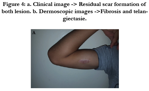
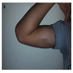
Figure 4: a. Clinical image -> Residual scar formation of both lesion. b. Dermoscopic images ->Fibrosis and telangiectasie.
Cutaneous tuberculosis with sporotrichoid pattern is exceptional, it may be observed in young people following post-traumatic inoculation [1]. Typically the lesions have alinear (lymphatic) arrangement as a result of lymphatic spread of the infection from a primary entry focus in the skin. The unique clinical presentation of the cutaneous tuberculosisshouldbe kept in mind in the differential diagnosis of sporotrichosis-like skin lesions, especially in elderly immuno-compromised patients [2]. Dermoscopy is a noninvasive diagnostic tool that recently has been discovered to assist also the clinical diagnosis of infectious skin diseases. However, dermosocopy of cutaneous tuberculosis is less reported and concerned mainly the lupus vulgaris, with typically the yellow to golden-colored background and linear vessels [3]. New features were revealed in our case, such as peripheral hairpin vessels and whitish streaks. this observation requires further confirmation on a larger series of cases of cutaneous tuberculosis. Cutaneous tuberculosis mimicking sporotrichosisis rare, the diagnosis remains difficult and must be based on clinical-dermoscopic and histological arguments. Nevertheless, it must be mentioned before any sporotrichoidlesion especially in an endemic country such as Morocco.
References
- Nakamura S, Hashimoto Y, Nishi K, Takahashi H, Takeda K, Mizumoto T, et al. Cutaneous tuberculosis simulating lymphocutaneous sporotrichosis. Australasian Journal of Dermatology. 2012 Nov;53(4):316-7.
- Akhdari N, Zouhair K, Habibeddine S, Lakhdar H. Tuberculose cutanée de l'enfant au Maroc: étude de 30 cas. Archives de pédiatrie. 2006 Aug 1;13(8):1098-101.
- Brasiello M, Zalaudek I, Ferrara G, Gourhant JY, Capoluongo P, Roma P, Argenziano G. Lupus vulgaris: a new look at an old symptom-the lupoma observed with dermoscopy. Dermatology. 2009;218(2):172 – 174.


